. 2017 Jun;23(11-12):572-584.
doi: 10.1089/ten.TEA.2016.0457. Epub 2017 Apr 14.

Cell and Growth Factor-Loaded Keratin Hydrogels for Treatment of Volumetric Muscle Loss in a Mouse Model

Affiliations

Cell and Growth Factor-Loaded Keratin Hydrogels for Treatment of Volumetric Muscle Loss in a Mouse Model

H B Baker et al. Tissue Eng Part A. 2017 Jun.

Abstract

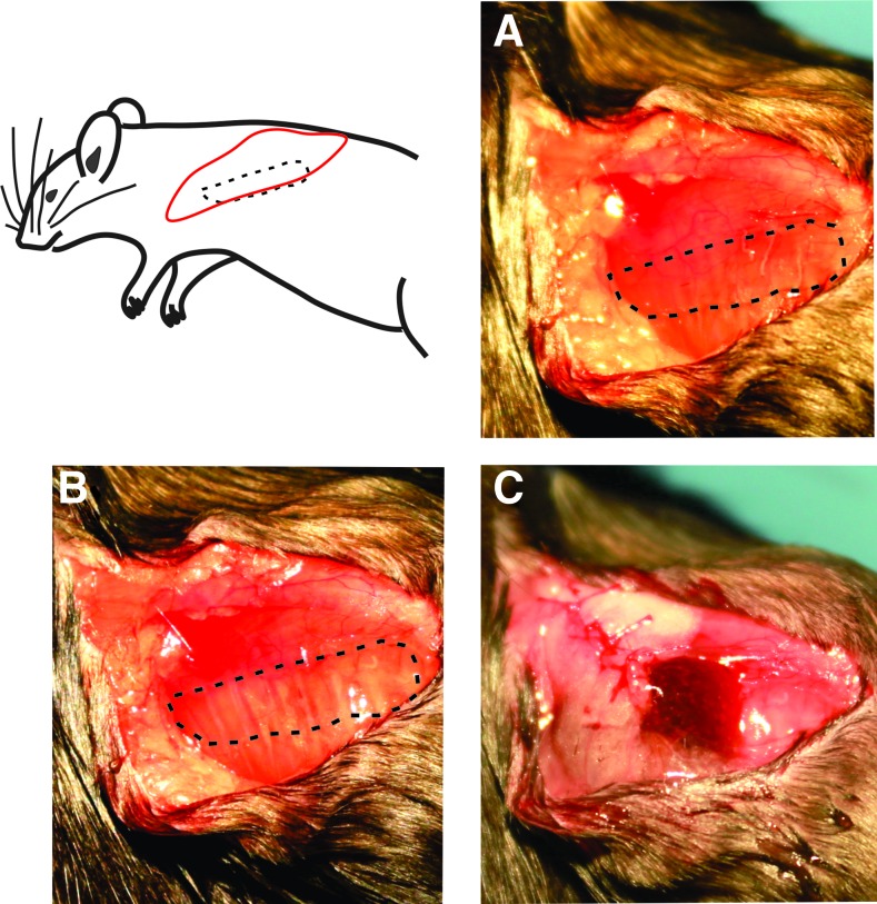
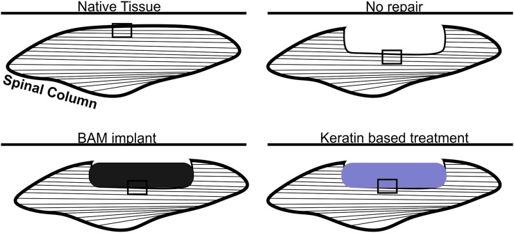
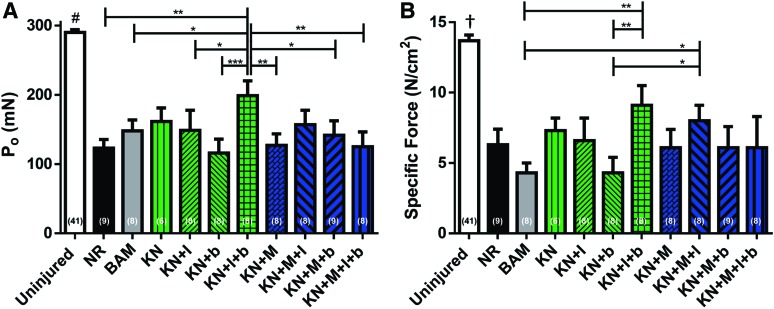
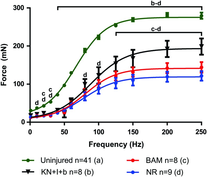
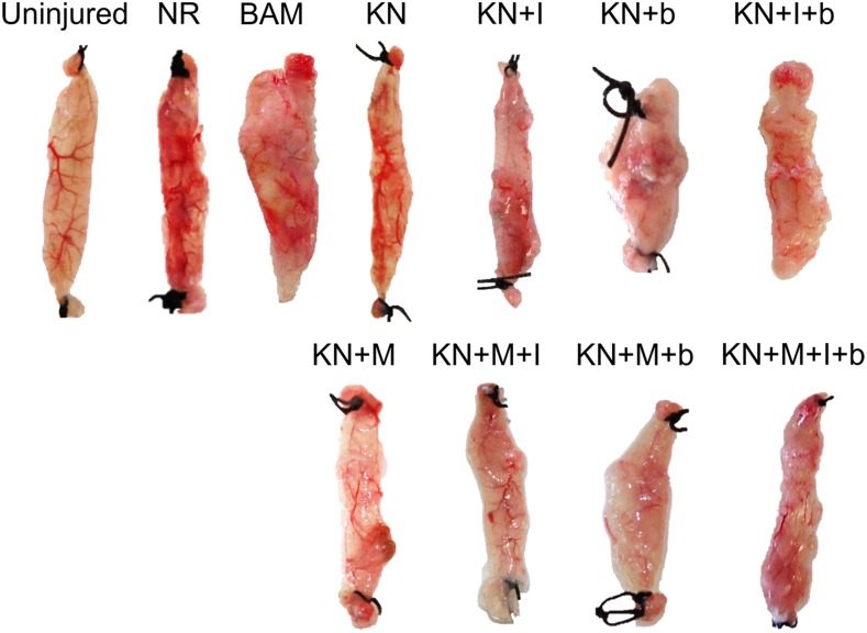
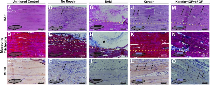
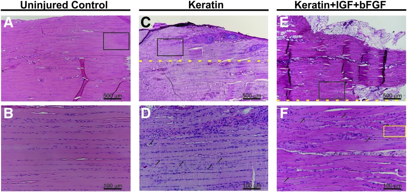
Wounds to the head, neck, and extremities have been estimated to account for ∼84% of reported combat injuries to military personnel. Volumetric muscle loss (VML), defined as skeletal muscle injuries in which tissue loss results in permanent functional impairment, is common among these injuries. The present standard of care entails the use of muscle flap transfers, which suffer from the need for additional surgery when using autografts or the risk of rejection when cadaveric grafts are used. Tissue engineering (TE) strategies for skeletal muscle repair have been investigated as a means to overcome current therapeutic limitations. In that regard, human hair-derived keratin (KN) biomaterials have been found to possess several favorable properties for use in TE applications and, as such, are a viable candidate for use in skeletal muscle repair. Herein, KN hydrogels with and without the addition of skeletal muscle progenitor cells (MPCs) and/or insulin-like growth factor 1 (IGF-1) and/or basic fibroblast growth factor (bFGF) were implanted in an established murine model of surgically induced VML injury to the latissimus dorsi (LD) muscle. Control treatments included surgery with no repair (NR) as well as implantation of bladder acellular matrix (BAM). In vitro muscle contraction force was evaluated at two months postsurgery through electrical stimulation of the explanted LD in an organ bath. Functional data indicated that implantation of KN+bFGF+IGF-1 (n = 8) enabled a greater recovery of contractile force than KN+bFGF (n = 8)***, KN+MPC (n = 8)**, KN+MPC+bFGF+IGF-1 (n = 8)**, BAM (n = 8)*, KN+IGF-1 (n = 8)*, KN+MPCs+bFGF (n = 9)*, or NR (n = 9)**, (*p < 0.05, **p < 0.01, ***p < 0.001). Consistent with the physiological findings, histological evaluation of retrieved tissue revealed much more extensive new muscle tissue formation in groups with greater functional recovery (e.g., KN+IGF-1+bFGF) when compared with observations in tissue from groups with lower functional recovery (i.e., BAM and NR). Taken together, these findings further indicate the general utility of KN biomaterials in TE and, moreover, specifically highlight their potential application in the treatment of VML injuries.

Keywords: FGF; IGF; keratin hydrogel; myogenesis; volumetric muscle loss.


Conflict of interest statement

No competing financial interests exist.

Figures

<b>FIG. 1.</b>
FIG. 1.
Surgical flow of volumetric muscle loss injury and keratin implant. Muscle is isolated in (A) with the proposed defect region outlined; defect is created and outlined in (B). Fascia is partially closed and keratin hydrogel is injected into the fascial pocket (C). Fascia and skin are then closed and the animal is allowed to recover. Color images available online at www.liebertpub.com/tea
<b>FIG. 2.</b>
FIG. 2.
Depiction of location of tissue sections for use in histological analyses. Tissues were sectioned longitudinally in the same plane as the length of the LD muscles at 5 μm thickness. Images were taken at the regions indicated with rectangles in the figure to include the interface between uninjured and treated tissue with native tissue in the lower portion of each image. LD, latissimus dorsi. Color images available online at www.liebertpub.com/tea
<b>FIG. 3.</b>
FIG. 3.
Muscle function analysis at 2 months postsurgery. Maximum isometric contraction force (P0) as a result of electrical field stimulation (1–250 Hz, 0.2 ms, 30 V) (A); normalized to PCSA (B). For both measurements, uninjured contralateral control muscles n = 41, NR n = 9, BAM n = 8, KN n = 6, KN + I n = 8, KN + b n = 8, KN+I+b n = 8, KN + M n = 8, KN+M+I n = 8, KN+M+b n = 9, and KN+M+I+b n = 8. Acellular hydrogel treatment groups are colored green, cellularized hydrogel treatment groups are colored blue. Significant differences are denoted as follows: *p < 0.05, **p < 0.01, ***p < 0.001, † different from all other groups p < 0.001, and #different from all other groups p < 0.0001. NR, no repair; BAM, bladder acellular matrix. Color images available online at www.liebertpub.com/tea
<b>FIG. 4.</b>
FIG. 4.
Selected force–frequency curves with fitted dose–response curves. At 2 months postsurgery, contractions were elicited through electrical stimuli from 1 to 250 Hz as shown for uninjured, KN+I+b, BAM, and NR muscles. Dose–response data have been curve fit according to Equation (2). Curve fitting was used to determine EC50 and Hill Slope values shown in Table 2. Letters above group curves denote groups that were statistically different at a given data point; brackets indicate differences were observed for all points included in the span of the bracket; p < 0.05; a: Uninjured; b: KN+I+b; c: BAM; d: NR. Color images available online at www.liebertpub.com/tea
<b>FIG. 5.</b>
FIG. 5.
Representative tissue gross morphology. Images shown were taken following functional assessment carried out at 2 months postsurgery. Representative samples from each group are shown. Color images available online at www.liebertpub.com/tea
<b>FIG. 6.</b>
FIG. 6.
Representative examples of mouse LD cell and tissue morphology and functional protein expression. (A–C) Uninjured control, (D–F) NR, (G–I) BAM, (J–L) KN, and (M–O) KN+I+b. Cell and tissue morphology was examined through hematoxylin and eosin staining (A, D, G, J, M) wherein nuclei are stained blue-purple and cytoplasm and cellular proteins are stained pink. Collagen deposition was examined through Masson's trichrome staining (B, E, H, K, N), in which tissue stains red, collagen stains blue, and nuclei stain black. The presence of new muscle tissue formation within the defect and treatment site was evaluated by staining for myosin (MF-20) (C, F, I, L, O); (#) denotes collagen deposition; new muscle formation is indicated with black arrows; and yellow dashed lines indicate the interface between native and remodeling tissue. It is important to note that even in the case of relatively robust new muscle tissue formation, the interface between the newly formed skeletal muscle and the native tissue was quite readily identified by the greater deposition of extracellular collagen and/or adipose tissue, as well as the presence of smaller muscle fibers, which frequently exhibited branching profiles and centrally located nuclei (Fig. 7). Color images available online at www.liebertpub.com/tea
<b>FIG. 7.</b>
FIG. 7.
Histological evidence of newly regenerated muscle fibers in keratin and keratin+IGF+bFGF-treated muscles. Hematoxylin and eosin stain of representative samples from the uninjured control groups (A, B), KN-treated (C, D), and KN+I+b-treated groups (E, F) highlighting the skeletal muscle morphology observed in native and newly formed (regenerating) muscle tissue postimplantation. As shown, (B, D, F) represent the higher magnification image of black boxes in (A, C, E), respectively. Arrows denote examples of muscle fibers with centrally located nuclei in the KN implant region. The yellow box encircles a representative example of a branching fiber, although such fibers were identified throughout the implant region. Yellow dashed lines indicate the interface between native and regenerating/remodeling tissue. Color images available online at www.liebertpub.com/tea

Similar articles

Cited by

References

    1. Grogan B.F., and Hsu J.R.; Skeletal Trauma Research Consortium. Volumetric muscle loss. J Am Acad Orthop Surg 19, S35, 2011 - PubMed
    1. Covey D.C. Blast and fragment injuries of the musculoskeletal system. J Bone Joint Surg Am 84-A, 1221, 2002 - PubMed
    1. Masini B.D., Waterman S.M., Wenke J.C., Owens B.D., Hsu J.R., and Ficke J.R. Resource utilization and disability outcome assessment of combat casualties from operation Iraqi Freedom and operation enduring freedom. J Orthop Trauma 23, 261, 2009 - PubMed
    1. Owens B.D., Kragh J.F., Jr., Wenke J.C., Macaitis J., Wade C.E., and Holcomb J.B. Combat wounds in operation Iraqi freedom and operation enduring freedom. J Trauma 64, 295–299, 2008 - PubMed
    1. Thiele O.C., Seeberger R., Engel M., Freier K., and Hoffmann J. Development of the clinical use of distant flaps for head and neck reconstruction. J Craniomaxillofac Surg 42, 79, 2014 - PubMed

Publication types

MeSH terms

LinkOut - more resources