Review
. 2017 Apr 15:234:4-20.
doi: 10.1016/j.virusres.2017.01.026. Epub 2017 Feb 2.

Picornaviral polymerase structure, function, and fidelity modulation

Affiliations
Review

Picornaviral polymerase structure, function, and fidelity modulation

Olve B Peersen. Virus Res. .

Abstract

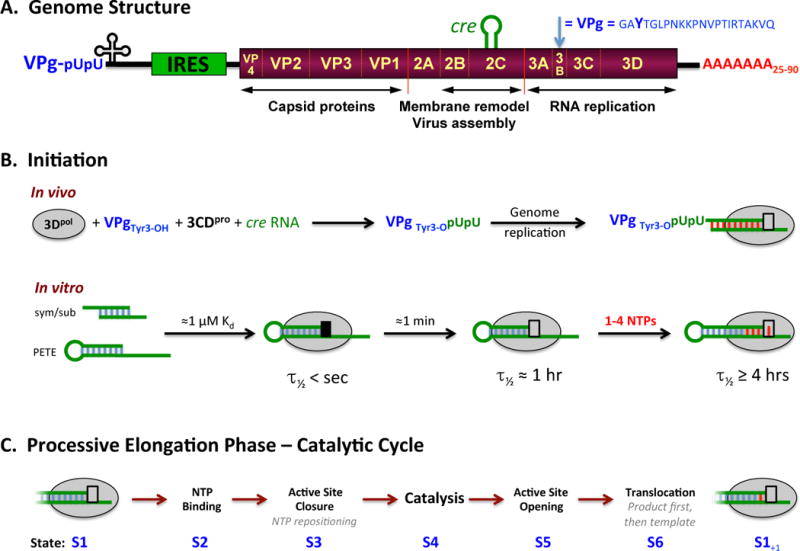
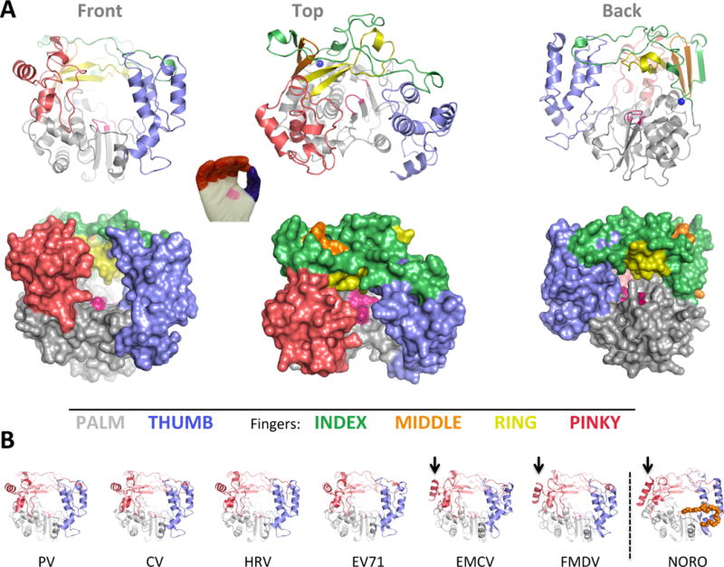
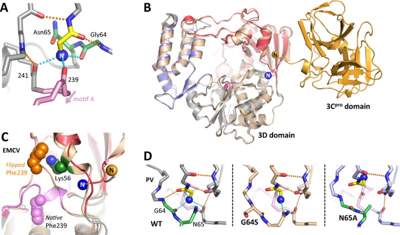
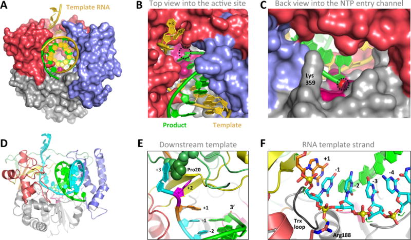
Like all positive strand RNA viruses, the picornaviruses replicate their genomes using a virally encoded RNA-dependent RNA polymerase enzyme known as 3Dpol. Over the past decade we have made tremendous advances in our understanding of 3Dpol structure and function, including the discovery of a novel mechanism for closing the active site that allows these viruses to easily fine tune replication fidelity and quasispecies distributions. This review summarizes current knowledge of picornaviral polymerase structure and how the enzyme interacts with RNA and other viral proteins to form stable and processive elongation complexes. The picornaviral RdRPs are among the smallest viral polymerases, but their fundamental molecular mechanism for catalysis appears to be generally applicable as a common feature of all positive strand RNA virus polymerases.

Keywords: Picornavirus; Polymerase; Positive strand RNA virus; RNA-dependent RNA polymerase; Structure.


Figures

Figure 1
Figure 1. Picornaviral Genome Structure and Polymerase Functions
(A) Schematic representation of the poliovirus genome as a representative picornavirus. The genome encodes a single ≈250 KDa polyprotein that is translated from an internal ribosome entry site (IRES) and cleaved into about a dozen smaller proteins and functional intermediates by the viral 2Apro, 3Cpro, and 3CDpro proteases. The last part of the polyprotein is 3Dpol, a RNA-dependent RNA polymerase that is only active upon cleavage of the 3Cpro–3Dpol junction. Many picornaviruses replace 2Apro with a N-terminal leader (L) protease. (B) The native initiation pathway for 3Dpol uses the viral VPg protein (i.e. 3B) whose Tyr3 becomes doubly uridylylated via a cre RNA templated reaction in the context of a viral replication center. In vitro, however, 3Dpol will initiate using short RNA duplexes, such as the self-complementary sym/sub sequences used extensively by the Cameron lab or RNA hairpins “PETE” constructs used by the Peersen group. There is a stepwise assembly pathway whereby the initial 3Dpol-RNA complex needs to undergo a conformational transition to become catalytically competent, as indicated by the black versus grey box at the active site. (C) The full catalytic cycle that takes place repeatedly during processive elongation can be divided into six major structural states, S1–S6, as previously described (Gong and Peersen, 2010).
Figure 2
Figure 2. Overview of picornaviral RdRP structures
(A) Cartoon and surface representations of poliovirus 3Dpol in three different orientations. The structure resembles a cupped right hand composed of palm, fingers, and thumb domains. The fingers domain can be further divided into five distinct structures (per color key), and the active site in the palm domain is shown as a patch of magenta. Note that the index finger reaches across the palm to contact the top of the thumb, creating a channel at the back of the enzyme whereby NTPs access the active site. (B) All the picornaviral 3Dpol structures solved to date exhibit a very high degree of structural homology. Note that one helix on the pinky finger changes orientation between the enteroviruses and EMCV/FMDV groups, with the latter resembling the helix orientation seen in the non-picornaviral norovirus 3Dpol. Norovirus polymerase also has a C-terminal extension (orange) that reaches into the RNA exit channel.
Figure 3
Figure 3. Proteolytic activation of 3Dpol
The very N-terminus (blue sphere N) of a picornaviral polymerase is buried in a pocket at the base of the fingers domain, resulting in activation of the enzyme through stabilization of the motif A movements needed for catalysis. (A) Structural interactions involving the PV 3Dpol N-terminus as viewed from the direction of the active site. (B) Backside view of the PV 3CDpro structure where the 3Dpol N-terminus does not exist because it is part of the flexible linker between the 3Cpro and 3Dpol domains. (C) In one EMCV 3Dpol structure [4YNZ] the N-terminus was no longer bound in its pocket and there is a major rearrangement of Phe239 within motif A. (D) Structural plasticity of the PV N-terminus binding pocket, where the native conformation is stabilized by six hydrogen bonds to residues Gly1 and Glu2. In the G64S structure one only of the native H-bonds to the N-terminus exists, but Asn65 has formed a new H-bond. Mutating Asn65 to alanine causes the pocket to collapse and the N-terminus folds up into the fingers domain.
Figure 4
Figure 4. RNA interactions with 3Dpol in the Elongation Complex
(A–C) Three views of the PV 3Dpol-RNA elongation complex oriented as in Figure 2A. The single stranded template RNA enters the polymerase from the top and takes a ≈90° turn as it contacts the palm at the active site, where it forms a duplex with the product strand and exits the polymerase via the wide front channel. The location of the priming 3′ hydroxyl group at the active site is marked with a dashed circle. (D) Cartoon representation of the elongation complex with the template strand in cyan and the fingers colored as in Figure 2A. (E) Details of key interaction within the 3Dpol-RNA complex, including Pro20 inserted between the +2 and +3 template strand bases, the binding pocket for the unstacked +2 nucleotide on the template strand, and the pre-positioning of the templating +1 nucleotide above the active site where it is poised for base pairing with an incoming NTP. (E) The conformation of the template RNA strand as it passes through the polymerase active site. Note the backbone linkage between the −1 and −2 nucleotides is not standard A-form (red arrow vs green arrows) due to a salt bridge interaction with Arg188.
Figure 5
Figure 5. Active site closure via a unique palm domain based mechanism
(A) The picornaviral RdRPs use the same two metal catalytic mechanism as other replicative polymerases, but close their active sites for catalysis by a novel movement of motif A in response to correct NTP binding. This moves the essential Asp233 into the active site to enable catalysis. (B) Structure of the closed PV 3Dpol active site following CTP binding and catalysis [3OL7], with the set of stabilizing hydrogen bonds directly linking the NTP 2′ hydroxyl to Asp238 in the repositioned motif A highlighted in magenta. (C) Structural details of the open and closed EV71 polymerase active sites where all twelve magnesium ion coordination ligands have been captured in a single structure. In the pre-catalysis open state the NTP phosphate interacts with Arg174 from the ring finger via two water molecules, and Arg174 is itself positioned by interactions with Glu161. The metal A magnesium ion is located ≈5 Å away from the active site in the open state, but is moved into the catalytic center during active site closure when both Asp233 and Asp329 reorient to support catalysis. In the closed state, the magnesiums are axially coordinated by the two Asp328 oxygens from below and by a pair of water molecules (W) from above.
Figure 6
Figure 6. RNA Translocation Mechanism
Following catalysis the 3Dpol active site opens back up without RNA translocation, unlike what seen in many other replicative polymerases where those two events are coupled. EV71 elongation complex structure suggest a Brownian ratchet mechanism for translocation. (A) Comparison of EV71 structures captured at the beginning state 1 [5F8L] and ending state 6 [5F8N] of a single nucleotide incorporation cycle show a sliding movement of the product RNA strand. In state 1 (top) the priming duplex is strained and its base pairs are not planar because the 3′ end of the product strand is being pulled toward the active site. After catalysis in state 6 (bottom) the product strand has moved toward the thumb such that the duplex now forms proper planar base pairs. Note that the template strand backbone does not move, as per the grey visual alignment lines. (B) Direct comparison of the base pair hydrogen bonding patterns at the beginning (lighter) and end (darker) of the catalytic cycle show how the product strand slides relative to the fixed template strand. (C) The movement toward the 5′ end is facilitate by the priming −1 nucleotide (blue) moving up and out the active site after the addition of the +1 nucleotide (yellow), releasing RNA-polymerase contacts and allowing the RNA to relax to the more favorable base pairing conformation.
Figure 7
Figure 7. Structures of 3Dpol–VPg complexes
(A) The structure of CV 3Dpol onto which the backbone structures of CV, EV71, and FMDV VPg complexes have been superimposed. The N- and C- ends of the VPg peptides are indicated in blue and red, respectively, and the parts of the EV71 VPg structure in weak electron density are shown in lighter purple. Note that only the FMDV VPg is bound near the active site. (B,C) Detailed views of FMDV VPg (and VPg-pU bounds near the active site, with the product RNA strand from PV elongation complex superimposed for comparison. Note the VPg Tyr3 OH group is bound above and across the active site from the normal RNA 3′ OH group and the uracil base of the VPg-pU complex is perpendicular to the bases of the normal RNA binding orientation. (D) Recombination mutants in PV 3Dpol shown on the wildtype EC structure in two orientations. The D79H mutation is located on the exterior surface of the polymerase far from the RNA binding surface. L420A, on the other hand, makes a direct contact with the product strand RNA at the third nucleotide from the active site (magenta) and biochemical data indicate it reduces recombination by reducing the efficiency of re-initiation on a new template strand.
Figure 8
Figure 8. Fidelity modulation sites in 3Dpol
Work from multiple groups over the past decade has identified several 3Dpol regions where mutations can affect replication fidelity and virus growth. (A) Structure of the poliovirus elongation complex showing the three major regions for fidelity mutants; 1: G64S, M296I, and H273R near the N-terminus, 2: motif A in dark purple, and 3: motif D with K359 and F363 in dark green. (B) Structural changes on the underside of motif A as it changes form the open (light) to the closed (dark) conformation during active site closure. Note the rotamer switch of Phe232 that allows the backbone to move significantly with minimal movement of the side chain itself. (C) Comparison of the N-terminus binding pocket structures of wildtype and M296I FMDV 3Dpol. The M296I variant is a higher fidelity polymerase, much like G64S in PV, and it has similar effects on the structure of the N-terminus even though the mutation is in the protein interior. (D) The structure and dynamics of the extensive hydration and hydrogen bonding network within the fingers domain involving His273 are likely affected by the low fidelity PV H273R mutation. (E) The sliding movement of Phe363 atop the motif D helix during active site closure, where the motif D loop is pulled in toward the active site along with motif A (purple). Mutating Phe363 to tryptophan increased 3Dpol fidelity. The motif D loop also contains T362 that is mutated to an isoleucine in the Sabin type 1 oral poliovirus strain, yielding a lower fidelity polymerase.

Similar articles

Cited by

References

    1. Agudo R, Ferrer-Orta C, Arias A, de la Higuera I, Perales C, Perez-Luque R, Verdaguer N, Domingo E. A multi-step process of viral adaptation to a mutagenic nucleoside analogue by modulation of transition types leads to extinction-escape. PLoS pathogens. 2010;6:e1001072. - PMC - PubMed
    1. Andino R, Domingo E. Viral quasispecies. Virology. 2015:479–480. - PMC - PubMed
    1. Arias A, Arnold JJ, Sierra M, Smidansky ED, Domingo E, Cameron CE. Determinants of RNA-dependent RNA polymerase (in)fidelity revealed by kinetic analysis of the polymerase encoded by a foot-and-mouth disease virus mutant with reduced sensitivity to ribavirin. J Virol. 2008;82:12346–12355. - PMC - PubMed
    1. Arnold JJ, Bernal A, Uche U, Sterner DE, Butt TR, Cameron CE, Mattern MR. Small ubiquitin-like modifying protein isopeptidase assay based on poliovirus RNA polymerase activity. Analytical biochemistry. 2006;350:214–221. - PMC - PubMed
    1. Arnold JJ, Cameron CE. Poliovirus RNA-dependent RNA polymerase (3D(pol)). Assembly of stable, elongation-competent complexes by using a symmetrical primer-template substrate (sym/sub) J Biol Chem. 2000;275:5329–5336. - PubMed

LinkOut - more resources