. 2017 Jan 20:10:127.
doi: 10.3389/fnana.2016.00127. eCollection 2016.

Dendritic Arborization Patterns of Small Juxtaglomerular Cell Subtypes within the Rodent Olfactory Bulb

Affiliations

Dendritic Arborization Patterns of Small Juxtaglomerular Cell Subtypes within the Rodent Olfactory Bulb

Wolfgang G Bywalez et al. Front Neuroanat. .

Abstract

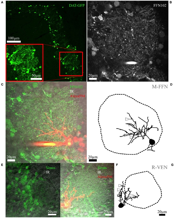
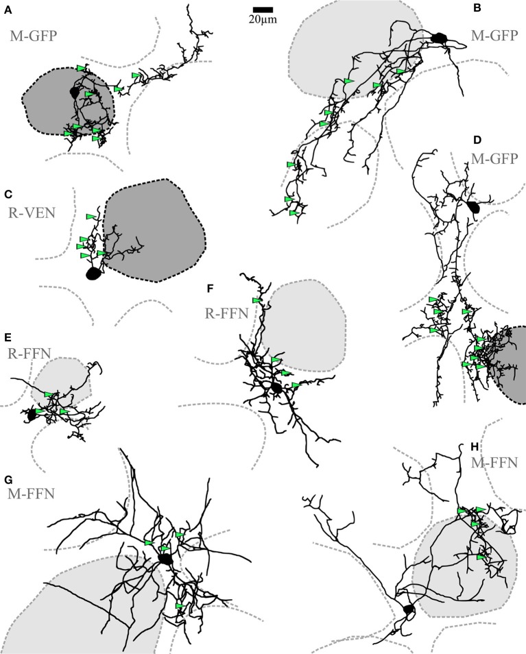
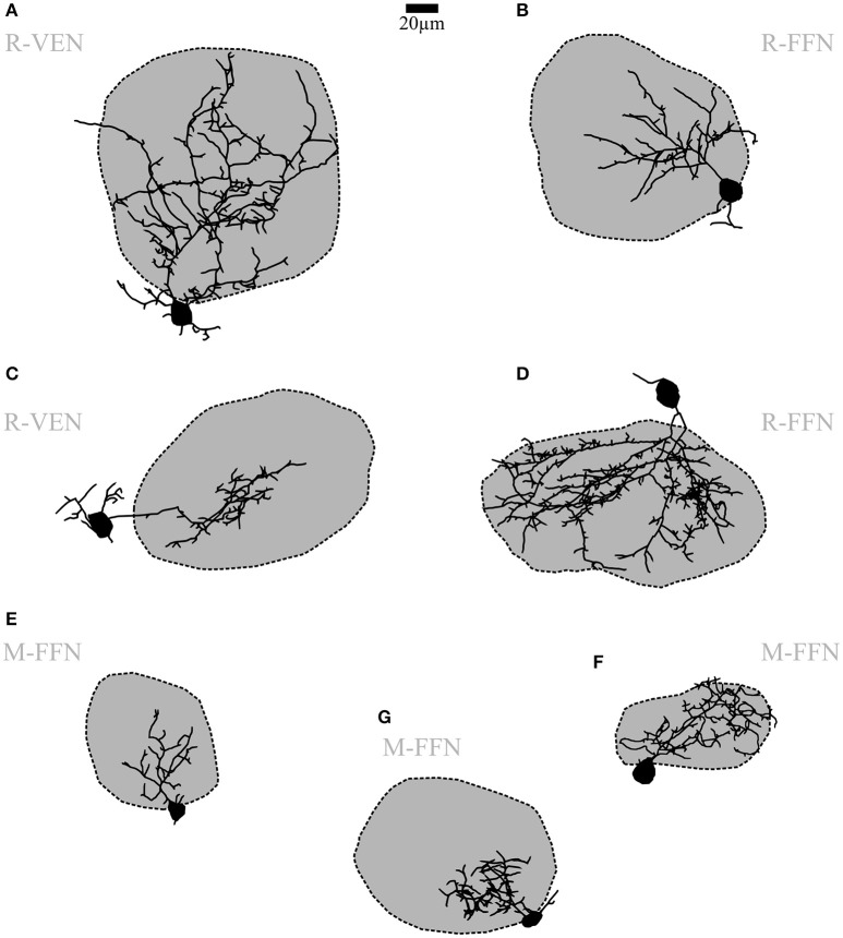
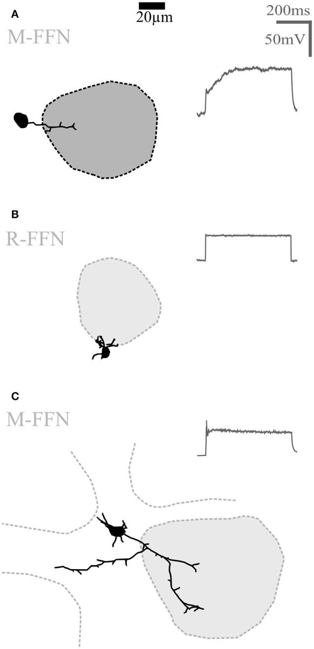
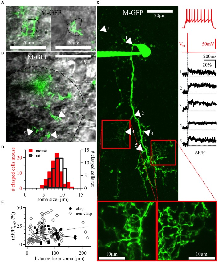
Within the glomerular layer of the rodent olfactory bulb, numerous subtypes of local interneurons contribute to early processing of incoming sensory information. Here we have investigated dopaminergic and other small local juxtaglomerular cells in rats and mice and characterized their dendritic arborization pattern with respect to individual glomeruli by fluorescent labeling via patching and reconstruction of dendrites and glomerular contours from two-photon imaging data. Dopaminergic neurons were identified in a transgenic mouse line where the expression of dopamine transporter (DAT) was labeled with GFP. Among the DAT+ cells we found a small short-axon cell (SAC) subtype featuring hitherto undescribed dendritic specializations. These densely ramifying structures clasped mostly around somata of other juxtaglomerular neurons, which were also small, non-dopaminergic and to a large extent non-GABAergic. Clasping SACs were observed also in wild-type mice and juvenile rats. In DAT+ SAC dendrites, single backpropagating action potentials evoked robust calcium entry throughout both clasping and non-clasping compartments. Besides clasping SACs, most other small neurons either corresponded to the classical periglomerular cell type (PGCs), which was never DAT+, or were undersized cells with a small dendritic tree and low excitability. Aside from the presence of clasps in SAC dendrites, many descriptors of dendritic morphology such as the number of dendrites and the extent of branching were not significantly different between clasping SACs and PGCs. However, a detailed morphometric analysis in relation to glomerular contours revealed that the dendrites of clasping SACs arborized mostly in the juxtaglomerular space and never entered more than one glomerulus (if at all), whereas most PGC dendrites were restricted to their parent glomerulus, similar to the apical tufts of mitral cells. These complementary arborization patterns might underlie a highly complementary functional connectivity. The morphometric approach may serve to differentiate also other subtypes of juxtaglomerular neurons, help to identify putative synaptic partners and thus to establish a more refined picture of glomerular network interactions during odor sensing.

Keywords: calcium imaging; dendrites; dendritic arborization analysis; dopaminergic neurons; juxtaglomerular cells; morphological reconstruction; subcellular compartments; two-photon imaging.


Figures

Figure 1
Figure 1
Fluorescent cell labeling and reconstructions. (A) Maximal z-projection scan of an OB slice from an adult mouse with tamoxifen-induced DAT-GFP+ cells surrounding the glomeruli. The inset shows a rescan at higher magnification. (B) Representative live FFN102 labeling of the glomerular and juxtaglomerular neuropil in an adult WT mouse brain slice. Green fluorescence channel of the TPLSM, with some brightly stained puncta within the glomerular neuropil as well as some labeled cell bodies at the glomerulus borders. (C) Same region in the trans-infrared detection (trans-IR) channel shown in gray overlaid with the green FFN102 fluorescence from (B) and the Alexa-594 stained, FFN102+ single neuron in red. (D) Maximal z-projection of the fully reconstructed cell from (C), including an outline of the glomerulus. The open arrowheads indicate exemplary spines; not all spines are labeled for sake of clarity. The selected cell is the PGC also depicted in Figure 3B. Similar representations of cells and glomeruli are also used in Figures 2–4. (E) Representative VGAT-Venus labeling of the glomerular and juxtaglomerular neuropil in a juvenile rat in the green and the trans-IR channel. (F) Another VGAT-Venus example overlaid with the trans-IR channel shown in gray and the Alexa-594 stained, Venus+ single neuron in red. (G) The respective reconstruction of the clasping cell and its parent glomerulus from (F) which is also shown in Figure 2C. The open arrowheads indicate exemplary spines; not all spines are labeled for sake of clarity.
Figure 2
Figure 2
Clasping cells (CCs). (A–H) Maximal z-projections of reconstructions of individual representative CCs with indicated surrounding glomeruli. The “parent glomerulus” is shaded for each cell (see methods). Darker shades indicate that the cell arborizes within the glomerular neuropil (A,C,D), whereas light shades indicate non-innervated parent glomeruli. Non-shaded structures are surrounding glomeruli, which the dendrites do not ramify in and which do not constitute the parent glomerulus. Green arrowheads indicate the position of clasped JGC somata. Most dendritic structures including cell body clasps do not ramify in the intraglomerular neuropil, with the exception of a few superficially entering branches, also including dense clasp-like bundles (A,C,D). R-VEN, VGAT-Venus+ cells in juvenile rats; R-FFN, FFN102+ cells in juvenile rats; M-FFN, FFN102+ cells in adult mice; M-GFP, DAT-GFP+ cells in adult mice.
Figure 3
Figure 3
Periglomerular cells (PGCs). (A–G) Maximal z-projections of reconstructions of individual representative PGCs with their corresponding contacted glomeruli. The dark shading indicates that dendrites contact the intraglomerular neuropil. Note the differing fractions of intraglomerular arborization with sparse or no juxtaglomerular processes, as well as the differing degree of branching and spine density across individual cells. R-VEN, VGAT-Venus+ cells in juvenile rats; R-FFN, FFN102+ cells in juvenile rats; M-FFN, FFN102+ cells in adult mice. Scale same size as in Figure 2.
Figure 4
Figure 4
Undersized cells (UCs). Maximal z-projections of reconstructions of representative UCs. Shading of glomeruli corresponding to Figures 2, 3. None of the reconstructed UCs showed truncations or signs of bad filling. UCs also never showed regular sodium action potential firing (voltage traces shown on the right in panels A–C) upon step depolarizations. (A) Morphologically very simple cell entering a single glomerulus. (B) Even smaller cell that does not enter the adjacent glomerulus. (C) Slightly larger cell compared to (A,B), extending dendrites in the juxtaglomerular space. R-FFN, FFN102+ cells in juvenile rats; M-FFN, FFN102+ cells in adult mice.
Figure 5
Figure 5
DAT+ clasping cell dendrites: Clasp structures and AP-evoked Ca2+ signals. (A) Two different clasping structures around cell bodies (left and right), with a dotted line indicating the somatic outline of the clasped cell—both structures are from a 20 weeks old DAT-GFP mouse (cell shown in panel C, the left clasping structure corresponds to the arrowhead number 4). (B) Exemplary, tight clasping structures (labeled “c”) within the glomerular neuropil (DAT-GFP mouse, 17 weeks old), with a dotted line indicating the glomerular border. (A,B) Show single z-plane scans from the green channel overlaid with the trans-IR channel in gray. (C) Maximal z-projection of the OGB-1 filled CC from Figure 2D (insets at bottom: magnified rescans of clasping structures). Numbered arrows in the scan correspond to the locations of the numbered averaged ΔF/F transients on the right, time-locked to the evoked somatic action potential (red trace directly above the black ΔF/F traces). The voltage recording on the top right shows this cell's accommodating firing pattern upon step depolarization (+90 pA current injection). In the scan, the arrow labeled with “a” points out a putative stained axon. It is thinner than the other, presumably dendritic, structures and bears no visible spines or clasps. (D) Distribution of the size of somata surrounded by clasps in juvenile rats (n = 45 clasped somata) and adult mice (both WT and DAT-GFP; n = 79 clasped somata). (E) Individual ΔF/F measurements versus the distance of their dendritic location from the soma (in n = 11 DAT-GFP+ mouse CCs; within clasps: black dots, n = 45 locations; within non-clasping dendrites: gray diamonds, n = 110 locations). No significant correlations emerged. M-GFP, DAT-GFP+ cells in adult mice.
Figure 6
Figure 6
Fractional glomerular arborization of clasping and periglomerular cells. (A) Schema of the three analyzed parent glomerulus' volumes: an outer shell comprising directly adjacent juxtaglomerular cell bodies (thickness 15 μm; green), an inner shell (thickness 15% of the mean glomerular radius; yellow) and a core region (red; see methods). (B) Fractional arborization patterns of the PGCs and CCs [and additional rat mitral cells (MCs)]: Intraglomerular dendritic voxel number divided by the outer shell number for all pooled cells (left), or separated by species (middle and right). (C) Fractional arborization of the same cell types in the combined shell volume (inner + outer shell), relative to the total juxtaglomerular and glomerular volume (shells + core), again pooled (left) or separated by species (middle and right). **P < 0.01; ***P < 0.005, Mann-Whitney-Test.

Similar articles

Cited by

References

    1. Adam Y., Livneh Y., Miyamichi K., Groysman M., Luo L., Mizrahi A. (2014). Functional transformations of odor inputs in the mouse olfactory bulb. Front. Neural Circuits 8:129. 10.3389/fncir.2014.00129 - DOI - PMC - PubMed
    1. Adam Y., Mizrahi A. (2011). Long-term imaging reveals dynamic changes in the neuronal composition of the glomerular layer. J. Neurosci. 31, 7967–7973. 10.1523/jneurosci.0782-11.2011 - DOI - PMC - PubMed
    1. Banerjee A., Marbach F., Anselmi F., Koh M. S., Davis M. B., Garcia da Silva P., et al. . (2015). An interglomerular circuit gates glomerular output and implements gain control in the mouse olfactory bulb. Neuron 87, 193–207. 10.1016/j.neuron.2015.06.019 - DOI - PMC - PubMed
    1. Batista-Brito R., Close J., Machold R., Fishell G. (2008). The distinct temporal origins of olfactory bulb interneuron subtypes. J. Neurosci. 28, 3966–3975. 10.1523/jneurosci.5625-07.2008 - DOI - PMC - PubMed
    1. Blackman A. V., Grabuschnig S., Legenstein R., Sjostrom P. J. (2014). A comparison of manual neuronal reconstruction from biocytin histology or 2-photon imaging: morphometry and computer modeling. Front Neuroanat. 8:65. 10.3389/fnana.2014.00065 - DOI - PMC - PubMed

LinkOut - more resources