

Hearing Impairment Is Associated with Smaller Brain Volume in Aging
- PMID: 28163683
- PMCID: PMC5247429
- DOI: 10.3389/fnagi.2017.00002
Hearing Impairment Is Associated with Smaller Brain Volume in Aging
Erratum in
-
Corrigendum: Hearing Impairment Is Associated with Smaller Brain Volume in Aging.Front Aging Neurosci. 2017 May 8;9:131. doi: 10.3389/fnagi.2017.00131. eCollection 2017. Front Aging Neurosci. 2017. PMID: 28491034 Free PMC article.
Abstract
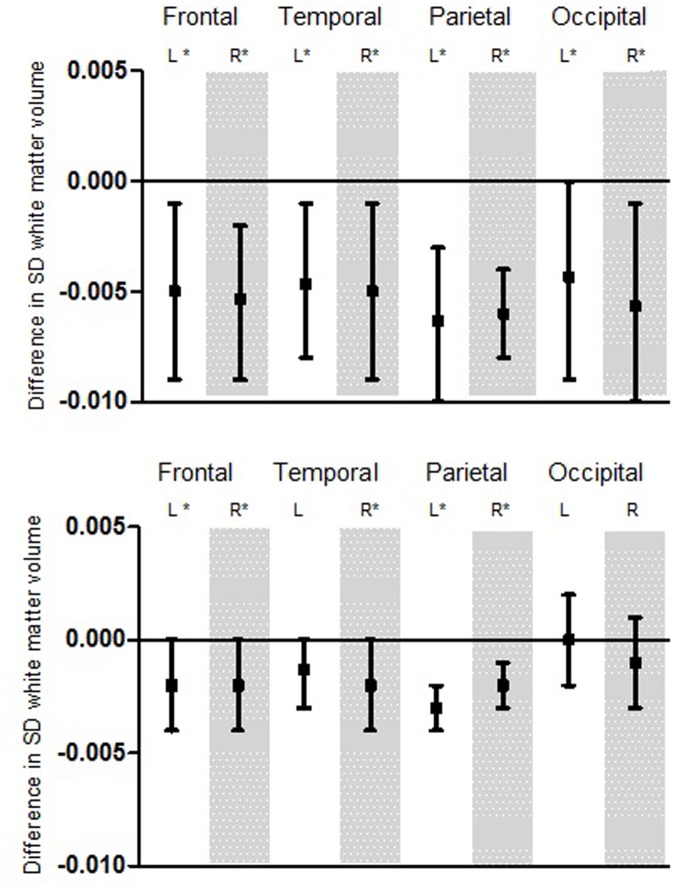
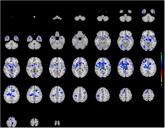
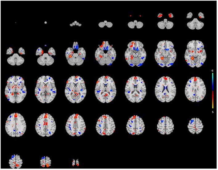
Although recent studies show that age-related hearing impairment is associated with cerebral changes, data from a population perspective are still lacking. Therefore, we studied the relation between hearing impairment and brain volume in a large elderly cohort. From the population-based Rotterdam Study, 2,908 participants (mean age 65 years, 56% female) underwent a pure-tone audiogram to quantify hearing impairment. By performing MR imaging of the brain we quantified global and regional brain tissue volumes (total brain volume, gray matter volume, white matter (WM) volume, and lobe-specific volumes). We used multiple linear regression models, adjusting for age, sex, head size, time between hearing test and MR imaging, and relevant cognitive and cardiovascular covariates. Furthermore, we performed voxel-based morphometry to explore sub-regional differences. We found that a higher pure-tone threshold was associated with a smaller total brain volume [difference in standardized brain volume per decibel increase in hearing threshold in the age-sex adjusted model: -0.003 (95% confidence interval -0.004; -0.001)]. Specifically, WM volume was associated. Both associations were more pronounced in the lower frequencies. All associations were consistently present in all brain lobes in the lower frequencies and in most lobes in the higher frequencies, and were independent of cognitive function and cardiovascular risk factors. In voxel-based analyses we found associations of hearing impairment with smaller white volumes and some smaller and larger gray volumes, yet these were statistically non-significant. Our findings demonstrate that hearing impairment in elderly is related to smaller total brain volume, independent of cognition and cardiovascular risk factors. This mainly seems to be driven by smaller WM volume, throughout the brain.
Keywords: age-related hearing impairment; brain MRI; pure-tone audiogram; voxel-based analysis; white matter.
Figures
References
-
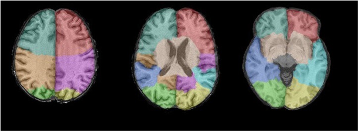
- Bokde A. L., Teipel S. J., Schwarz R., Leinsinger G., Buerger K., Moeller T. (2005). Reliable manual segmentation of the frontal, parietal, tempora land occipital lobes on magnetic resonance images of healthy subjects. Brain Res. Brain Res. Protoc. 14 135–145. 10.1016/brainresprot.2004.10.001 - DOI - PubMed
LinkOut - more resources
Full Text Sources
Other Literature Sources