. 2017 Jan 20:8:07.
doi: 10.3389/fphar.2017.00007. eCollection 2017.

The New Synthetic H2S-Releasing SDSS Protects MC3T3-E1 Osteoblasts against H2O2-Induced Apoptosis by Suppressing Oxidative Stress, Inhibiting MAPKs, and Activating the PI3K/Akt Pathway

Affiliations

The New Synthetic H2S-Releasing SDSS Protects MC3T3-E1 Osteoblasts against H2O2-Induced Apoptosis by Suppressing Oxidative Stress, Inhibiting MAPKs, and Activating the PI3K/Akt Pathway

Xiaofei Yan et al. Front Pharmacol. .

Abstract

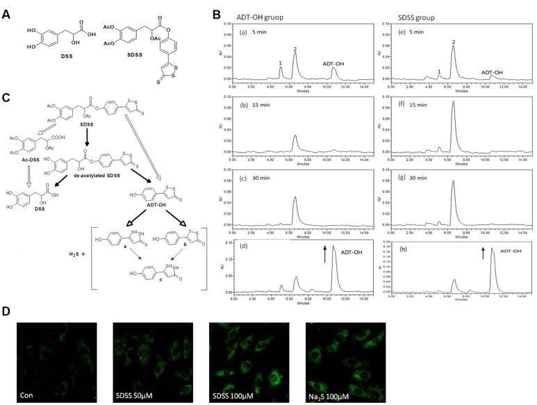
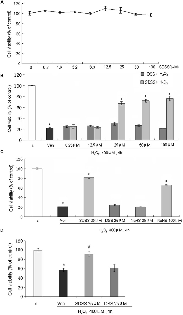
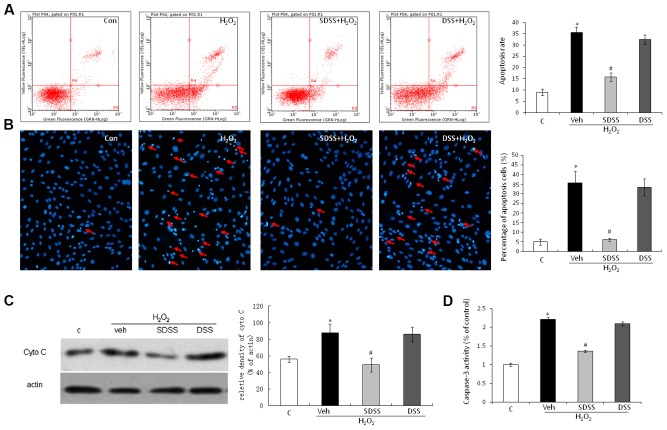
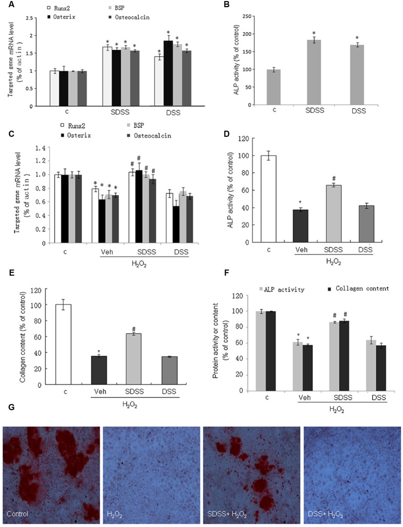
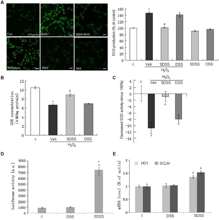
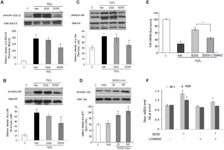
Reactive oxygen species (ROS) are important in osteoporosis development. Oxidative stress induces apoptosis of osteoblasts and arrest of their differentiation. Both Danshensu (DSS) and hydrogen sulfide (H2S) produce significant antioxidant effect in various systems. In this study, we synthesized SDSS, a novel H2S-releasing compound derived from DSS, and studied its antioxidant effect in an H2O2-induced MC3T3-E1 osteoblastic cell injury model. We first characterized the H2S releasing property of SDSS in both in vivo and in vitro models. HPLC chromatogram showed that intravenous injection of SDSS in adult rats released ADT-OH, a well proved H2S sustained-release moiety, within several minutes in the rat plasma. Using an H2S selective fluorescent probe, we further confirmed that SDSS released H2S in MC3T3-E1 osteoblastic cells. Biological studies revealed that SDSS had no significant toxic effect but produced protective effects against H2O2-induced MC3T3-E1 cell apoptosis. SDSS also reversed the arrest of cell differentiation caused by H2O2 treatment. This was caused by the stimulatory effect of SDSS on bone sialoprotein, runt-related transcription factor 2, collagen expression, alkaline phosphatase activity, and bone nodule formation. Further studies revealed that SDSS reversed the reduced superoxide dismutase activity and glutathione content, and the increased ROS production in H2O2 treated cells. In addition, SDSS significantly attenuated H2O2-induced activation of p38-, ERK1/2-, and JNK-MAPKs. SDSS also stimulated phosphatidylinositol 3-kinase/Akt signaling pathway. Blockade of this pathway attenuated the cytoprotective effect of SDSS. In conclusion, SDSS protects MC3T3-E1 cells against H2O2-induced apoptosis by suppressing oxidative stress, inhibiting MAPKs, and activating the phosphatidylinositol 3-kinase/Akt pathway.

Keywords: MAPK signaling; PI3K/AKT; hydrogen sulfide donating drugs; osteoblast; reactive oxygen species.


Figures

FIGURE 1
FIGURE 1
Characterization of the new H2S releasing compound SDSS. (A) Structural formulas of DSS and SDSS. SDSS is a hybrid of Ac-DSS and ADT-OH. (B) HPLC chromatograms of SDSS. Rats were administered with 30 mg/kg ADT-OH (a-c) or SDSS (e–g). Chromatograms were recorded at 330 nm by analyzing plasma 5, 15, and 30 min after injection. For the bottom panels (d and h), samples from ADT-OH (d) and SDSS (h) groups were spiked with additional solution of ADT-OH in ACN (with the arrow) and immediately analyzed by HPLC. The final concentrations of spiking ADT-OH in sample ADT-OH and SDSS were 0.01 mM and 0.02 mM, respectively. (C) Proposed diagram of metabolic degradation of SDSS and the release of H2S. The metabolic degradation of AFT-OH was proposed by Giustarini et al. (2010a). (D) Detection of intracellular H2S released from SDSS in MC3T3-E1 cells. Cells were pretreated with probe 8 (20 μM) in DOTAP liposome for 2 h, and then treated with SDSS or Na2S for 3 h. Representative fluorescent images were taken using a confocal microscopy. Ex = 633 nm, Em above 650 nm. Magnification: ×200. Both Na2S and SDSS significantly increased the fluorescence intensity.
FIGURE 2
FIGURE 2
Effects of SDSS on H2O2-induced cell injury in MC3T3-E1 osteoblastic cells. Cell viability was determined by the methylthiazolyltetrazolium assay. (A) Effect of SDSS (0–100 μM) alone. (B) Concentration-dependent effect of SDSS and DSS against H2O2-induced cell injury. (C) Comparison of the effect of DSS, SDSS, and NaHS on H2O2-treated cells (400 μM, 4 h). (D) Effect of SDSS on viability of primary cultured rat osteoblasts in cells treated with H2O2 for 4 h. Mean ± standard error of the mean, n = 4–8. p < 0.05 versus the control groups (c); #p < 0.05 versus the H2O2 groups; veh, vehicle.
FIGURE 3
FIGURE 3
Effect of SDSS on H2O2-induced cell apoptosis. (A) Cell apoptosis was detected by annexin V–fluorescein isothiocyanate/propidium iodide double staining and examined with a fluorescence-activated cell sorting flow cytometry analyzer. (B) Cell apoptosis was detected by Hoechst 33342 staining in cells treated with vehicle, H2O2, H2O2+ DSS (25 μM) and H2O2 + SDSS (25 μM). Cells with condensed or fragmented nuclei were identified as apoptosis cells and counted based on nuclear condensation or fragmentation. (C) Representative Western blots and group data for cytochrome c cytosolic expression. Pretreatment with SDSS (25 μM, 1 h) significantly inhibited H2O2 induced release of cytochrome c. (D) Caspase 3 activity in differently treated cells. SDSS reduced the H2O2-induced increased caspase 3 activity. Mean ± standard error of the mean, n = 4–8. p < 0.05 versus control (c); #p < 0.05 versus H2O2 group; cyto, cytochrome; DSS, danshensu; veh, vehichle.
FIGURE 4
FIGURE 4
Effect of SDSS on MC3T3-E1 cell differentiation. (A,C) Quantitative reverse transcription PCR analysis of the messenger RNA (mRNA) levels of bone sialoprotein (BSP), runt-related transcription factor 2 (Runx2), osterix, and osteocalcin in the absence (A) or presence (C) of H2O2. (B,D) Effect of SDSS and DSS on alkaline phosphatase (ALP) activity in cells treated without (B) and with (D) H2O2. (E) SDSS but not DSS attenuated H2O2 down-regulated collagen expression in MC3T3-E1 cells on day 5 after H2O2 treatment. (F) Effect of SDSS and DSS on ALP activity and collagen expression in the primary cultured osteoblasts on day 5 after H2O2 treatment. (G) Representative image of mineralized bone nodule formation. H2O2 decreased bone nodule formation and SDSS alleviated the effect of H2O2. Mean ± standard error of the mean, n = 4-8. p < 0.05 versus control (c); #p < 0.05 versus H2O2 group.
FIGURE 5
FIGURE 5
Effect of SDSS on the production of reactive oxygen species (ROS) and the activities of antioxidants. (A) Representative images (left) and group data (right) showing that SDSS, but not DSS, suppressed H2O2-induced ROS production. (B,C) SDSS, but not DSS, reversed H2O2-reduced GSH (B) and SOD (C) activity. (D,E) SDSS, but not DSS, stimulated antioxidant response element-luciferase activity (D) and quantitative reverse transcription PCR analysis of the messenger RNA (mRNA) level of heme oxygenase 1 (HO1) and glutamate-cysteine ligase modifier subunit (GCLM) (E). Mean ± standard error of the mean, n = 4–8. p < 0.05 versus the control group; #p < 0.05 versus the H2O2 group; c, control; DSS, danshensu; Veh, vehicle group.
FIGURE 6
FIGURE 6
Involvement of mitogen-activated protein kinases and the phosphatidylinositol 3-kinase/Akt pathway in the protective effect of SDSS. Representative Western blots and group data showing the effect of SDSS and DSS on H2O2-stimulated extracellular signal-regulated kinase 1/2 (Erk1/2; A), p38 (B), and c-Jun N-terminal kinase (Jnk; C) phosphorylation. (D) Representative Western blots and group data showing that SDSS (25 and 100 μM) induced Akt phosphorylation. (E,F) LY294002 (15 μM, 15 min) attenuated the effect of SDSS (25 μM, 18 h) on cell viability (E), and messenger RNA (mRNA) expression levels of heme oxygenase 1 (HO-1) and glutamate-cysteine ligase modifier subunit (GCLM) (F). Mean ± standard error of the mean, n = 4–6. p < 0.05 versus control group; #p < 0.05 versus the H2O2 group; +p < 0.05 versus the SDSS group; c, control; DSS, danshensu; Veh, vehicle group.

Similar articles

Cited by

References

    1. Bai X. C., Lu D., Bai J., Zheng H., Ke Z. Y., Li X. M., et al. (2004). “Oxidative stress inhibits osteoblastic differentiation of bone cells by ERK and NF-kappaB. Biochem. Biophys. Res. Commun. 314 197–207. 10.1016/j.bbrc.2003.12.073 - DOI - PubMed
    1. Banerjee Mustafi S., Chakraborty P. K., Dey R. S., Raha S. (2009). Heat stress upregulates chaperone heat shock protein 70 and antioxidant manganese superoxide dismutase through reactive oxygen species (ROS), p38MAPK, and Akt. Cell Stress Chaperones 14 579–589. 10.1007/s12192-009-0109-x - DOI - PMC - PubMed
    1. Basu S., Michaelsson K., Olofsson H., Johansson S., Melhus H. (2001). Association between oxidative stress and bone mineral density. Biochem. Biophys. Res. Commun. 288 275–279. 10.1006/bbrc.2001.5747 - DOI - PubMed
    1. Cooper C. (1999). Epidemiology of osteoporosis. Osteoporos. Int. 9(Suppl. 2), S2–S8. 10.1007/PL00004156 - DOI - PubMed
    1. Cui G., Shan L., Hung M., Lei S., Choi I., Zhang Z., et al. (2013). A novel Danshensu derivative confers cardioprotection via PI3K/Akt and Nrf2 pathways. Int. J. Cardiol. 168 1349–1359. 10.1016/j.ijcard.2012.12.012 - DOI - PubMed