Review
. 2017 Jan 20:8:5.
doi: 10.3389/fphys.2017.00005. eCollection 2017.

The Multifaceted Role of SNARE Proteins in Membrane Fusion

Affiliations
Review

The Multifaceted Role of SNARE Proteins in Membrane Fusion

Jing Han et al. Front Physiol. .

Abstract

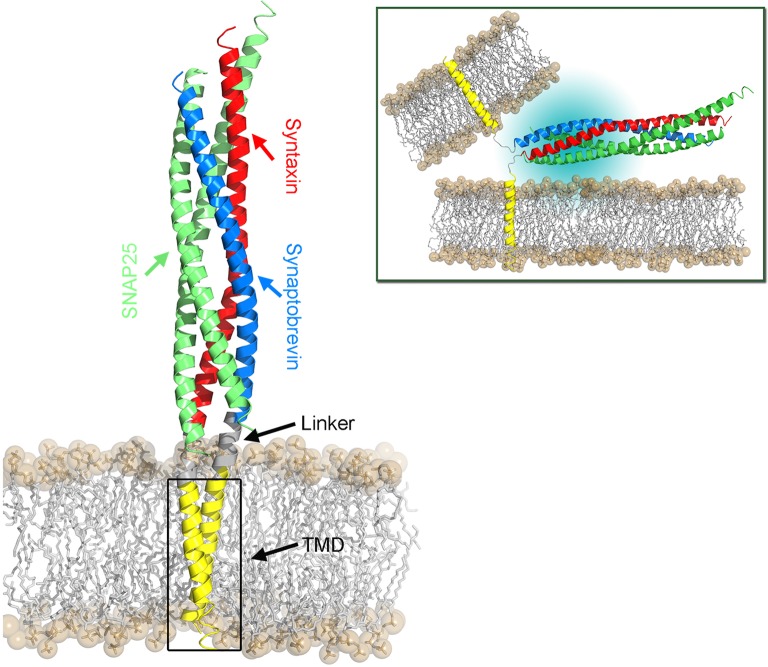
Membrane fusion is a key process in all living organisms that contributes to a variety of biological processes including viral infection, cell fertilization, as well as intracellular transport, and neurotransmitter release. In particular, the various membrane-enclosed compartments in eukaryotic cells need to exchange their contents and communicate across membranes. Efficient and controllable fusion of biological membranes is known to be driven by cooperative action of SNARE proteins, which constitute the central components of the eukaryotic fusion machinery responsible for fusion of synaptic vesicles with the plasma membrane. During exocytosis, vesicle-associated v-SNARE (synaptobrevin) and target cell-associated t-SNAREs (syntaxin and SNAP-25) assemble into a core trans-SNARE complex. This complex plays a versatile role at various stages of exocytosis ranging from the priming to fusion pore formation and expansion, finally resulting in the release or exchange of the vesicle content. This review summarizes current knowledge on the intricate molecular mechanisms underlying exocytosis triggered and catalyzed by SNARE proteins. Particular attention is given to the function of the peptidic SNARE membrane anchors and the role of SNARE-lipid interactions in fusion. Moreover, the regulatory mechanisms by synaptic auxiliary proteins in SNARE-driven membrane fusion are briefly outlined.

Keywords: SNAP-25; SNARE; fusion regulation; membrane fusion; protein-lipid interactions; synaptobrevin; syntaxin.


Figures

Figure 1
Figure 1
Topology of the SNARE complex consisting of synaptobrevin (in blue), syntaxin-1A (in red), and two SNAP-25 (sn1 and sn2, both in green) proteins (PDB:1SFC, Sutton et al., 1998).
Figure 2
Figure 2
Schematic representation of a membrane fusion process following the stalk-intermediate pathway preceded by the docking stage during exocytosis. (A) Initial interaction between synaptobrevin (red) and syntaxin (violet) cytosolic domains. The energy released upon trans-SNARE complex formation is used to bring the membranes into close proximity (overcoming the repulsive forces between the negatively charged vesicles) and to partially dehydrate them. After SNARE complex formation the vesicles are in a “docked” state. (B) Upon triggering, nascent hydrophobic contacts between the approaching membranes are built between splaying lipids. (C) A stalk is subsequently formed and the lipids in the outer leaflets start to mix. (D) Stalk elongation leads to hemi-diaphragm (HD) formation and elongation. (E) Inner leaflets of opposing membranes begin to mix accompanied by pore formation. In the following, the pore expands until either one large vesicle is formed out of two small ones, or until all lipids from a small vesicle are fully incorporated into a planar target membrane.
Figure 3
Figure 3
Structural model of the SNARE complex embedded in a POPC lipid bilayer. The cis-SNARE complex (PDB:3IPD) at the post-fusion stage comprises two SNAP-25, one syntaxin, and one synaptobrevin protein. The latter two SNAREs consist of a cytoplasmic domain (SNARE motifs), a short linker domain, and a transmembrane domain (TMD). The bilayer head groups are shown as spheres and hydrophobic tails as sticks. The SNARE complex in the pre-fusion stage is shown in the top right panel. Here, the TMDs of synaptobrevin and syntaxin proteins are located in their respective host membranes.
Figure 4
Figure 4
Spatial sampling of the juxtamembrane region (JMR) in dependence of the primary TMD sequence of synaptobrevin. The left column shows a contour density plot of the JMR configurational sampling (center of mass positions) in the bilayer plane (x-y). The blue dot marks the position of the TMD. The right column shows the side view of the sampled conformational space of the juxtamembrane region (after fitting the TMD). In the right panel sampling of three copies of the peptide (sequence as shown in Table 1) is shown in different colors, namely in yellow, blue, and violet, respectively. Samplings were recorded in the time interval from 500 to 1000 ns of atomistic simulations (see Han et al., for details).
Figure 5
Figure 5
An alternative mechanism of fusion pore formation by the insertion of the TMDs into the membrane interior as suggested by Fang and Lindau (2014).
Figure 6
Figure 6
Structural model of synaptobrevin's most abundant TMD dimer embedded in a POPC bilayer (Han et al., 2015) as obtained from a sequential multiscaling approach (Pluhackova and Böckmann, 2015).
Figure 7
Figure 7
Structural models of sybII oligomers containing 3–8 copies of transmembrane domains (left to right). The sybII oligomer structures were obtained either from self-assembly CG-MD simulations (trimer and tetramer) or from manual built (pentamer to octamer), see Han et al. (2016a).
Figure 8
Figure 8
Lipid tail protrusion in a curved POPC membrane evoked by a sybII TMD peptide. Lipids are shown as gray sticks with phosphates highlighted as yellow spheres. The peptide is shown as a rainbow-colored cartoon and sticks. The protruding lipid is highlighted as thick sticks with carbons colored blue and oxygens red.
Figure 9
Figure 9
Structure of the Ca2+-bound synaptotagmin 1-SNARE complex (Zhou et al., 2015). SNAP25 shown in green, synaptobrevin in blue, syntaxin-1A in red, one copy of synaptotagmin-1 in violet and one in yellow.

Similar articles

Cited by

References

    1. Aeffner S., Reusch T., Weinhausen B., Salditt T. (2012). Energetics of stalk intermediates in membrane fusion are controlled by lipid composition. Proc. Natl. Acad. Sci. U.S.A. 109, E1609–E1618. 10.1073/pnas.1119442109 - DOI - PMC - PubMed
    1. Al-Abdul-Wahid M. S., DeMill C. M., Serwin M. B., Prosser R. S., Stewart B. A. (2012). Effect of juxtamembrane tryptophans on the immersion depth of synaptobrevin, an integral vesicle membrane protein. Biochim. Biophys. Acta Biomembranes 1818, 2994–2999. 10.1016/j.bbamem.2012.07.018 - DOI - PubMed
    1. Ammar M., Kassas N., Chasserot-Golaz S., Bader M. F., Vitale N. (2013). Lipids in regulated exocytosis: what are they doing? Front. Endocrinol. 4:125. 10.3389/fendo.2013.00125 - DOI - PMC - PubMed
    1. Bacia K., Schuette C. G., Kahya N., Jahn R., Schwille P. (2004). SNAREs prefer liquid-disordered over “raft” (liquid-ordered) domains when reconstituted into giant unilamellar vesicles. J. Biol. Chem. 279, 37951–37955. 10.1074/jbc.M407020200 - DOI - PubMed
    1. Bai J., Tucker W. C., Chapman E. R. (2004). PIP2 increases the speed of response of synaptotagmin and steers its membrane-penetration activity toward the plasma membrane. Nat. Struct. Mol. Biol. 11, 36–44. 10.1038/nsmb709 - DOI - PubMed

LinkOut - more resources