. 2017:2017:4651238.
doi: 10.1155/2017/4651238. Epub 2017 Jan 9.

Activin A Modulates CRIPTO-1/HNF4 α+ Cells to Guide Cardiac Differentiation from Human Embryonic Stem Cells

Affiliations

Activin A Modulates CRIPTO-1/HNF4 α+ Cells to Guide Cardiac Differentiation from Human Embryonic Stem Cells

Robin Duelen et al. Stem Cells Int. 2017.

Abstract

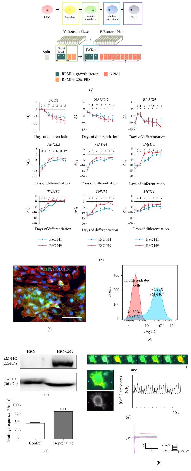
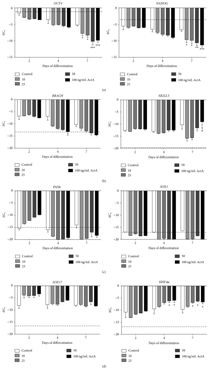
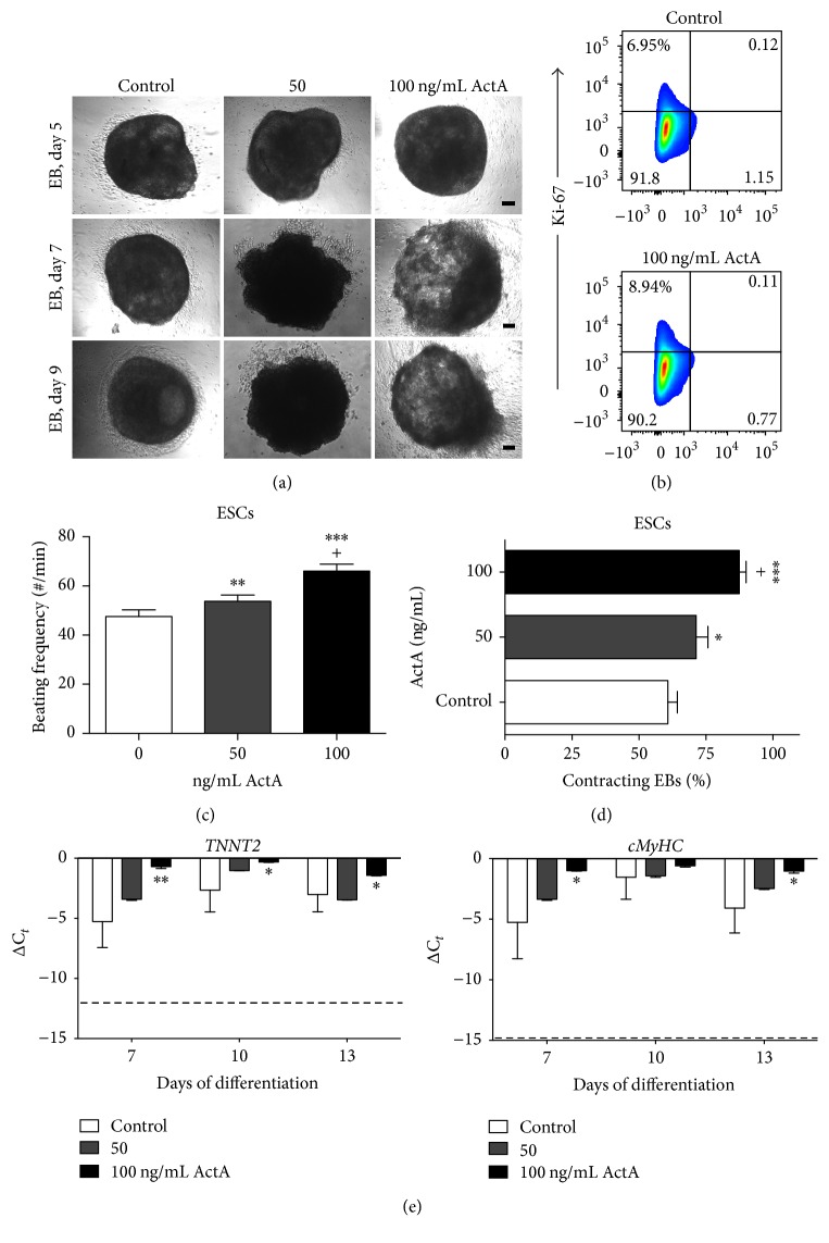
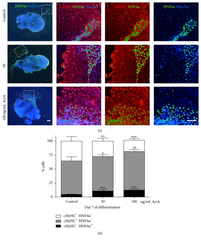
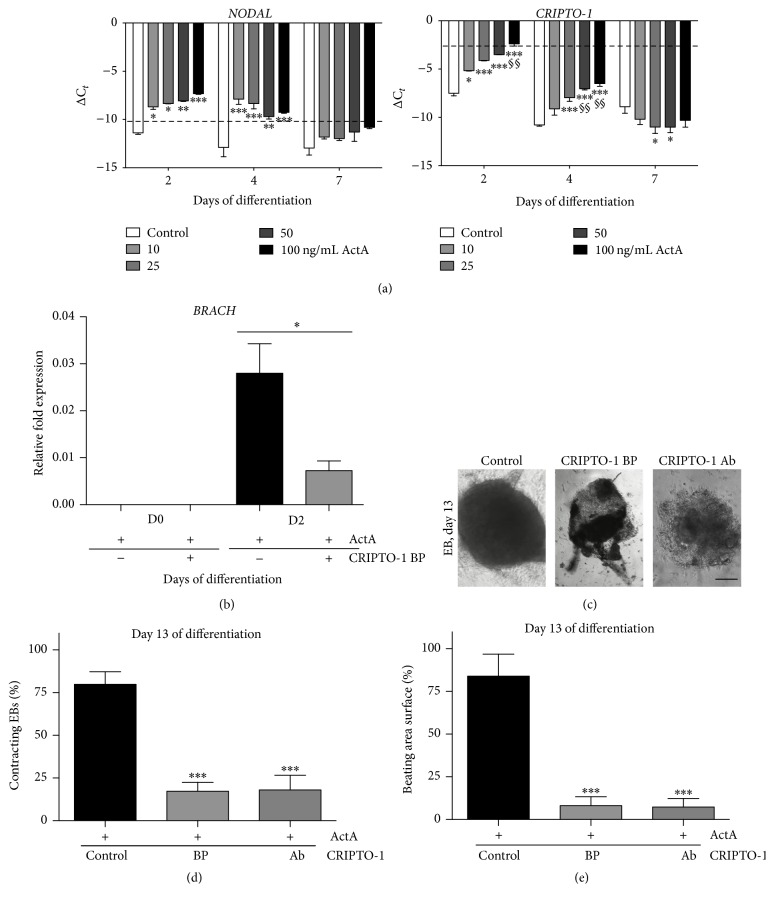
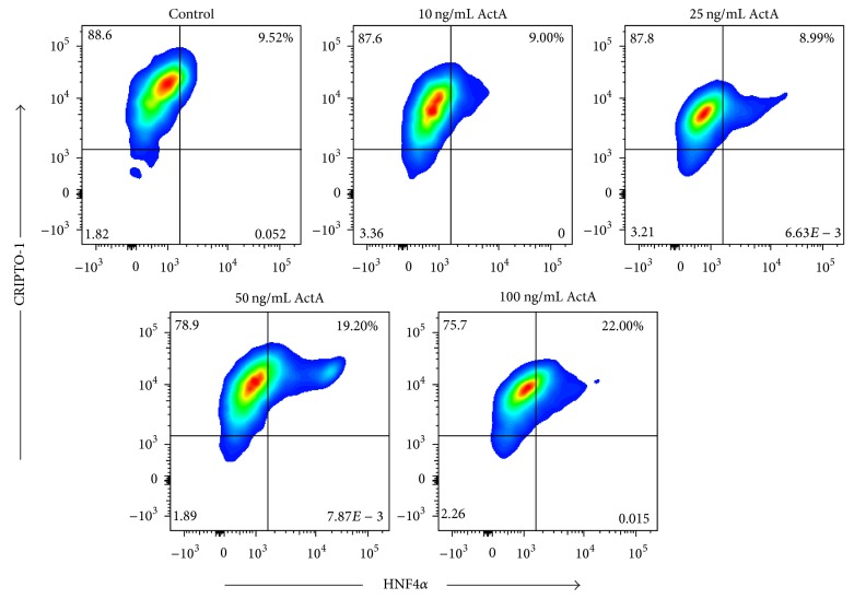
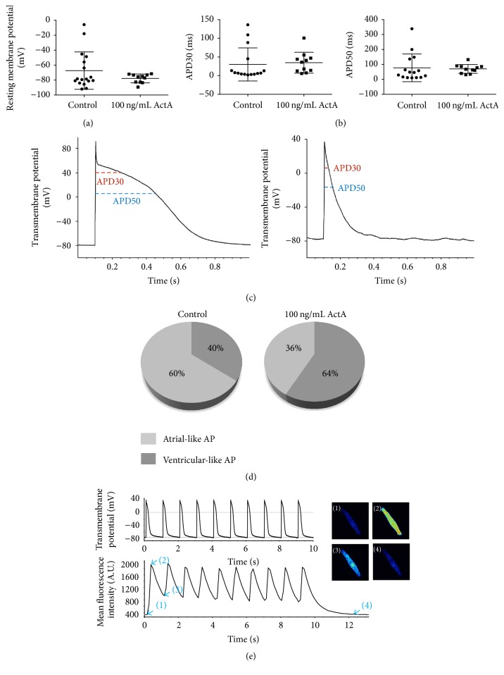
The use of human pluripotent stem cells in basic and translational cardiac research requires efficient differentiation protocols towards cardiomyocytes. In vitro differentiation yields heterogeneous populations of ventricular-, atrial-, and nodal-like cells hindering their potential applications in regenerative therapies. We described the effect of the growth factor Activin A during early human embryonic stem cell fate determination in cardiac differentiation. Addition of high levels of Activin A during embryoid body cardiac differentiation augmented the generation of endoderm derivatives, which in turn promoted cardiomyocyte differentiation. Moreover, a dose-dependent increase in the coreceptor expression of the TGF-β superfamily member CRIPTO-1 was observed in response to Activin A. We hypothesized that interactions between cells derived from meso- and endodermal lineages in embryoid bodies contributed to improved cell maturation in early stages of cardiac differentiation, improving the beating frequency and the percentage of contracting embryoid bodies. Activin A did not seem to affect the properties of cardiomyocytes at later stages of differentiation, measuring action potentials, and intracellular Ca2+ dynamics. These findings are relevant for improving our understanding on human heart development, and the proposed protocol could be further explored to obtain cardiomyocytes with functional phenotypes, similar to those observed in adult cardiac myocytes.


Conflict of interest statement

All the authors declare no potential conflict of interests.

Figures

Figure 1
Figure 1
Characterization of the embryoid body- (EB-) based cardiac differentiation protocol. Characterization was performed on human embryonic stem cell-derived cardiomyocytes (ESC-CMs) without the addition of Activin A (ActA) during the early stage of differentiation (control condition). (a) Schematic representation of the EB-based cardiac differentiation protocol. (b) Expression profiles for pluripotency (OCT4 and NANOG), mesodermal (BRACH), cardiac progenitor (NKX2.5 and GATA4), and late-stage CM (cMyHC, TNNT2, TNNI3, and HCN4) markers, monitored during 19 days of differentiation. (c) IF analyses of dissociated EBs at day 20 of differentiation, showing areas of connected CMs (cMyHC; red) expressing connexin 43 (CX43; green). Nuclei were stained with Hoechst (blue). (d) Flow cytometry analyses of cMyHC+ CMs at day 20 of differentiation. Representative example of three independent experiments. (e) Western blot analyses quantifying the cMyHC protein levels of undifferentiated ESCs and CMs after 20 days of differentiation, normalized by GAPDH. (f) β-Adrenergic response of CMs to isoprenaline (1 μM; isoproterenol), triggered at day 20 of differentiation. (g) Time course of intracellular Ca2+ handling at day 20 of differentiation, using the Ca2+-sensitive fluorescent indicator Fluo-4 (2.5 μM), monitored by confocal microscopy. (h) Whole-cell Na+ recording, accessed by applying 500 ms voltage pulses to potentials between −120 and +10 mV in 5 mV increments from a holding potential of −100 mV at 0.5 Hz. RT-QPCR data are represented as ΔCt, normalized for the housekeeping genes GAPDH, HPRT, and RPL13a. Data are representative of three independent experiments and values are expressed as mean ± standard error of the mean (SEM). Significant difference is versus control and indicated as P < 0.001: ∗∗∗. Scale bar = 100 μm.
Figure 2
Figure 2
Gene expression analyses of human embryonic stem cells (ESCs) subjected to cardiac differentiation in the presence of Activin A (ActA). Expression profiles of human ESC-derived CMs (ESC-CMs) for (a) pluripotency (OCT4, NANOG), (b) mesodermal (BRACH, NKX2.5), (c) ectodermal (PAX6, SOX1), and (d) endodermal (SOX17, HNF4α) lineage markers, monitored at days 0, 2, 4, and 7 of differentiation. Dashed lines show basal expression levels of undifferentiated ESCs. Each data point is represented as ΔCt, normalized for the housekeeping genes GAPDH, HPRT, and RPL13a. Note that ActA treatment during the early phase of cardiomyocyte (CM) differentiation directed human ESCs towards the mesendoderm cell fate. Data are representative of three independent experiments and values are expressed as mean ± standard error of the mean (SEM). Significant differences are indicated as P < 0.05: versus control, $ versus 10 ng/mL ActA, and # versus 25 ng/mL ActA; P < 0.01: ∗∗ versus control; P < 0.001: ∗∗∗ versus control.
Figure 3
Figure 3
ActA increased human ESC-CM differentiation efficiency. High doses of 50 and 100 ng/mL ActA promoted CM differentiation. (a) Brightfield images of EB morphology at days 5, 7, and 9 of differentiation of ActA treatment conditions (50 and 100 ng/mL) compared to the untreated cells (0 ng/mL ActA). (b) Proliferation was assessed using Ki-67, showing that 6.95% of the control cells and 8.94% of the ActA treated cells were in a proliferative stage. Flow cytometry analysis is a representative example of three independent experiments. (c) Quantification of contracting rate (beating frequency EBs per minute) and (d) percentage of contracting EBs at day 10 of differentiation. Efficiency is expressed as percentage of wells containing beating EBs to the total number of wells. (e) Gene expression analyses of late-stage CM genes (TNNT2, cMyHC) during 13 days of differentiation. Dashed lines show basal expression levels of undifferentiated ESCs. Each data point is represented as ΔCt, normalized for the housekeeping genes GAPDH, HPRT, and RPL13a. Data are representative of three independent experiments and expressed as mean ± SEM. Significant differences are indicated as P < 0.05: versus control and + versus 50 ng/mL; P < 0.01: ∗∗ versus control; P < 0.001: ∗∗∗ versus control. Scale bar = 100 μm.
Figure 4
Figure 4
ActA promoted CM differentiation by inducing HNF4α+ endoderm-like cell population. High doses of 50 and 100 ng/mL ActA during the early phases of cardiac differentiation increased the amount of HNF4α+ endoderm-like cells, which subsequently promoted the CM differentiation efficiency. (a) IF analyses for HNF4α (green), cMyHC (red), and Hoechst (blue) in 7-day-old EBs for control and 50 and 100 ng/mL ActA treated conditions. Dashed squares indicate the region of interest, corresponding to IF pictures at higher magnification. (b) Quantifications of cMyHC+, HNF4α+ and cMyHC HNF4α cell populations in the presence (50 and 100 ng/mL) or absence of ActA are reported as percentages of control conditions. Data are representative of three independent experiments and expressed as mean ± SEM. Significant differences are versus control and indicated as P < 0.05: ; P < 0.01: ∗∗; and P < 0.001: ∗∗∗. Scale bar = 100 μm.
Figure 5
Figure 5
Human ESC-CM differentiation potential after CRIPTO-1 interference. CRIPTO-1 blocking peptide (BP) impaired ActA-directed in vitro cardiac differentiation. (a) Gene expression of the TGF-β family members NODAL and CRIPTO-1 during a 13-day differentiation period. Dashed lines show basal expression levels of undifferentiated ESCs. (b) Normalized fold change of the mesodermal marker BRACH, measured at day 2 of differentiation after CRIPTO-1 BP treatment. Data, normalized for the housekeeping genes GAPDH, HPRT, and RPL13a, are representative of three independent experiments and expressed as mean ± SEM. (c) Brightfield pictures of ESC-CMs at day 13 of differentiation after CRIPTO-1 BP treatment or after addition of a human CRIPTO-1 antibody (Ab). (d) Percentage of contracting EBs and of (e) beating area surface in differentiated CMs pretreated with 100 ng/mL ActA and 5 µM CRIPTO-1 BP or pretreated with 100 ng/mL ActA and a human CRIPTO-1 Ab (10 µM). Data, normalized for the housekeeping genes GAPDH, HPRT, and RPL13a, are representative of four independent experiments and expressed as mean ± SEM. Significant differences are indicated as P < 0.05: versus control; P < 0.01: ∗∗ versus control and §§ versus 10 ng/mL ActA; P < 0.001: ∗∗∗ versus control. Scale bar = 200 μm.
Figure 6
Figure 6
CRIPTO-1 and HNF4α expression profiles in ActA treated human ESCs during early cardiac differentiation. HNF4α+ endodermal-like cells had a higher expression level of CRIPTO-1 in the control and 50 and 100 ng/mL ActA condition compared to the 10 and 25 ng/mL ActA treatment. Flow cytometry analyses of HNF4α and CRIPTO-1 expression after induction of human ESC-CM differentiation with the indicated concentrations of ActA. Representative example of three independent experiments.
Figure 7
Figure 7
Phenotyping CM differentiation showed both atrial- and ventricular-like (action potentials) APs. (a) Resting membrane potential (RMP) of human ESCs-CMs, measured during current clamp (whole-cell patch clamp recording) with or without a pretreatment of 100 ng/mL ActA. Each data point is a single cell measurement. (b) AP duration at 30% and 50% repolarization (APD30 and APD50). (c) Example of AP from a typical ventricular-like cell (left panel) and an atrial-like cell (right panel). (d) Percentages of CMs exhibiting an atrial-like or ventricular-like AP phenotype with and without ActA addition. (e) Simultaneous intracellular Ca2+ measurements using Fluo-4 and current clamp recording in a human ESC differentiated into CMs.

Similar articles

Cited by

References

    1. Burridge P. W., Keller G., Gold J. D., Wu J. C. Production of de novo cardiomyocytes: human pluripotent stem cell differentiation and direct reprogramming. Cell Stem Cell. 2012;10(1):16–28. doi: 10.1016/j.stem.2011.12.013. - DOI - PMC - PubMed
    1. Rajala K., Pekkanen-Mattila M., Aalto-Setälä K. Cardiac differentiation of pluripotent stem cells. Stem Cells International. 2011;2011:12. doi: 10.4061/2011/383709.383709 - DOI - PMC - PubMed
    1. Mummery C. L., Zhang J., Ng E. S., Elliott D. A., Elefanty A. G., Kamp T. J. Differentiation of human embryonic stem cells and induced pluripotent stem cells to cardiomyocytes: a methods overview. Circulation Research. 2012;111(3):344–358. doi: 10.1161/circresaha.110.227512. - DOI - PMC - PubMed
    1. Mummery C., Ward-van Oostwaard D., Doevendans P., et al. Differentiation of human embryonic stem cells to cardiomyocytes: role of coculture with visceral endoderm-like cells. Circulation. 2003;107(21):2733–2740. doi: 10.1161/01.cir.0000068356.38592.68. - DOI - PubMed
    1. Conlon F. L., Lyons K. M., Takaesu N., et al. A primary requirement for nodal in the formation and maintenance of the primitive streak in the mouse. Development. 1994;120(7):1919–1928. - PubMed

LinkOut - more resources