

Optic Nerve Sheath Meningocele: A Case Report
- PMID: 28163760
- PMCID: PMC5289384
- DOI: 10.3109/01658107.2013.766219
Optic Nerve Sheath Meningocele: A Case Report
Abstract
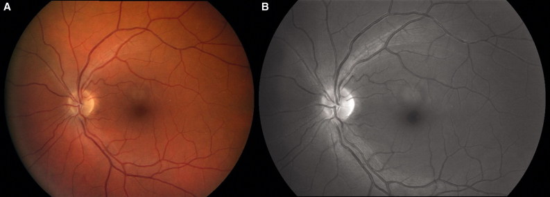
Isolated optic nerve sheath meningocele is a rare affection defined as the cystic enlargement of the optic nerve sheath filled with cerebrospinal fluid. We report the case of a 39-year-old woman presenting with bilateral meningocele uncovered during a routine examination for headache complaints. A 5-year follow-up validated the lesion's clinical and imaging stability. Magnetic resonance imaging (MRI) is an essential tool in the diagnosis of this pathology, alongside characteristic symptoms indicating that the meningocele might have progressively expanded into the orbit. In this case we present a therapeutic approach based on pathophysiological hypotheses and review of the literature.
Keywords: Optic nerve; optic nerve sheath meningocele; tear of the optic nerve sheath.
Figures


Similar articles
-
Case report: Bilateral optic nerve sheath meningocele: clinical aspects.Front Ophthalmol (Lausanne). 2024 May 8;4:1385485. doi: 10.3389/fopht.2024.1385485. eCollection 2024. Front Ophthalmol (Lausanne). 2024. PMID: 38984125 Free PMC article.
-
Surgically verified case of optic sheath nerve meningocele: case report with review of the literature.Neurosurg Rev. 1997;20(3):201-5. doi: 10.1007/BF01105565. Neurosurg Rev. 1997. PMID: 9297723 Review.
-
Stabilization of Visual Function After Optic Nerve Sheath Fenestration for Optic Nerve Meningocele.Ophthalmic Plast Reconstr Surg. 2017 Nov/Dec;33(6):e160-e161. doi: 10.1097/IOP.0000000000000932. Ophthalmic Plast Reconstr Surg. 2017. PMID: 28538611
-
Unilateral optic nerve sheath meningocele presented with amblyopia.J Pediatr Ophthalmol Strabismus. 2011 Aug 2;48 Online:e65-6. doi: 10.3928/01913913-20110726-03. J Pediatr Ophthalmol Strabismus. 2011. PMID: 21800781
-
Optic nerve sheath meningoceles. Clinical and radiographic features in 13 cases with a review of the literature.Ophthalmology. 1990 Nov;97(11):1519-31. Ophthalmology. 1990. PMID: 2255524 Review.
Cited by
-
Case report: Bilateral optic nerve sheath meningocele: clinical aspects.Front Ophthalmol (Lausanne). 2024 May 8;4:1385485. doi: 10.3389/fopht.2024.1385485. eCollection 2024. Front Ophthalmol (Lausanne). 2024. PMID: 38984125 Free PMC article.
-
A Pathophysiological Approach to Spontaneous Orbital Meningoceles: Case Report and Systematic Review.J Pers Med. 2024 Apr 28;14(5):465. doi: 10.3390/jpm14050465. J Pers Med. 2024. PMID: 38793047 Free PMC article. Review.
-
The relation of optic nerve sheath diameter (ONSD) and intracranial pressure (ICP) in pediatric neurosurgery practice - Part II: Influence of wakefulness, method of ICP measurement, intra-individual ONSD-ICP correlation and changes after therapy.Childs Nerv Syst. 2020 Jan;36(1):107-115. doi: 10.1007/s00381-019-04336-4. Epub 2019 Aug 8. Childs Nerv Syst. 2020. PMID: 31392457
-
Bilateral idiopathic optic nerve sheath meningocele: A case report and literature review.J Neurosci Rural Pract. 2022 Oct-Dec;13(4):781-784. doi: 10.25259/JNRP_5_2022. Epub 2022 Nov 2. J Neurosci Rural Pract. 2022. PMID: 36743743 Free PMC article.
References
-
- Garrity JA, Trautmann JC, Bartley GB, Forbes G, Bullock JD, Jones TW, Jr, Waller RR. Optic nerve sheath meningoceles. Clinical and radiographic features in 13 cases with a review of the literature. Ophthalmology 1990;97:1519–1531 - PubMed
-
- Lunardi P, Farah JO, Ruggeri A, Nardacci B, Ferrante L, Puzzilli F. Surgically verified case of optic sheath nerve meningocele: case report with review of the literature. Neurosurg Rev 1997;20:201–205 - PubMed
-
- Shanmuganathan V, Leatherbarrow B, Ansons A, Laitt R. Bilateral idopathic optic nerve sheath meningocele associated with unilateral transient cystoid macular oedema. Eye (Lond) 2002;16:800–802 - PubMed
-
- Hayreh SS. The sheath of the optic nerve. Ophthalmologica 1984;189:54–63 - PubMed
Publication types
LinkOut - more resources
Full Text Sources
Other Literature Sources