

Unilateral Optic Nerve Hypoplasia with Contralateral Optic Pathway Hypoplasia: A Case Report
- PMID: 28163766
- PMCID: PMC5289582
- DOI: 10.3109/01658107.2013.785572
Unilateral Optic Nerve Hypoplasia with Contralateral Optic Pathway Hypoplasia: A Case Report
Abstract
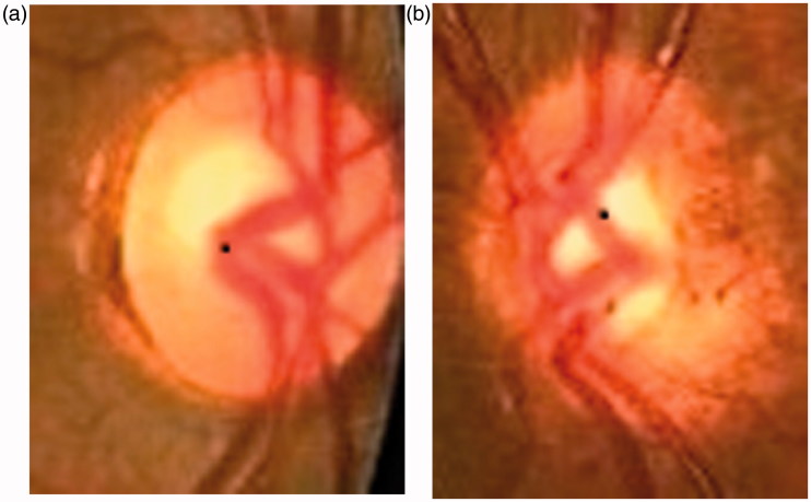
Optic nerve hypoplasia is diagnosed by the ophthalmoscopic appearance of the fundus of the eye and by standard magnetic resonance imaging of the brain. The ability to study eyes with optic nerve hypoplasia by magnetic resonance diffusion tensor imaging has improved the evaluation of the optic pathways. The authors report a case of unilateral optic nerve hypoplasia with hypoplasia of the contralateral optic pathway. The entire visual pathway of this patient was examined by magnetic resonance and magnetic resonance diffusion tensor imaging. The images show a decrease of the volume of the optic radiation contralateral to the optic nerve abnormality and also pre- and post-chiasmal abnormalities.
Keywords: Magnetic resonance diffusion tensor imaging; optic pathway hypoplasia; unilateral optic nerve hypoplasia.
Figures


Similar articles
-
Optic nerve hypoplasia. Identification by magnetic resonance imaging.Arch Ophthalmol. 1990 Nov;108(11):1562-7. doi: 10.1001/archopht.1990.01070130064032. Arch Ophthalmol. 1990. PMID: 2244841 Clinical Trial.
-
A case of amblyopia with contralateral optic nerve hypoplasia.Clin Exp Optom. 2013 Sep;96(5):500-3. doi: 10.1111/cxo.12007. Epub 2013 Jan 17. Clin Exp Optom. 2013. PMID: 23331175
-
Septo-optic dysplasia with unilateral optic nerve hypoplasia: case report.Chang Gung Med J. 2000 May;23(5):303-8. Chang Gung Med J. 2000. PMID: 10916232
-
Unilateral megalopapilla and contralateral optic nerve hypoplasia: a case report and review of the literature.J AAPOS. 2010 Feb;14(1):83-4. doi: 10.1016/j.jaapos.2009.10.007. Epub 2009 Dec 31. J AAPOS. 2010. PMID: 20045364 Review.
-
[Diffusion tensor imaging of the visual pathway in glaucomatous optic nerve atrophy].Ophthalmologe. 2017 Oct;114(10):906-921. doi: 10.1007/s00347-017-0467-1. Ophthalmologe. 2017. PMID: 28251307 Review. German.
References
-
- De Morsier G. Studies on malformation of cranio-encephalic sutures. Schweiz Arch Neurol Psychiatr 1956;77:267–292 - PubMed
-
- Barkovich AJ, Fram EK, Norman D. Septo-optic dysplasia: MR imaging. Radiology 1989;171:189–192 - PubMed
-
- Salmela MB, Cauley KA, Nickerson JP, Koski CJ, Flippi CG. Magnetic resonance diffusion tensor imaging (MRDTI) and tractography in children with septo-optic dysplasia. Pediatr Radiol 2010;40:708–713 - PubMed
-
- Taoka T, Sakamoto M, Nakagawa H, Nakase H. Diffusion tensor tractography of the Meyer loop in cases of temporal lobe resection for temporal lobe epilepsy: correlation between postsurgical visual field defect and anterior limit of Meyer loop on tractography. Am J Neuroradiol 2008;29:1329–1334 - PMC - PubMed
-
- Creel D, Wikop CJ, King RA. Asymmetric visually evoked potentials in human albinos; evidence for visual system anomalies. Invest Ophthalmol Visual Sci 1974;13:430–440 - PubMed
Publication types
LinkOut - more resources
Full Text Sources
Other Literature Sources