. 2017 Jan-Feb;11(1-2):E50-E57.
doi: 10.5489/cuaj.3789. Epub 2017 Jan 12.

Plasmacytoid urothelial carcinoma (PUC): Imaging features with histopathological correlation

Affiliations

Plasmacytoid urothelial carcinoma (PUC): Imaging features with histopathological correlation

Andrew D Chung et al. Can Urol Assoc J. 2017 Jan-Feb.

Abstract

Introduction: Plasmacytoid urothelial carcinoma (PUC) is a high-grade variant of conventional urothelial cell carcinoma. This study is the first to describe the imaging findings of PUC, which are previously unreported, using clinical and histopathological correlation.

Methods: With internal review board approval, we identified 22 consecutive patients with PUC from 2007-2014. Clinical parameters, including age, gender, therapy, surgical margins, and long-term outcome, were recorded. Baseline imaging was reviewed by an abdominal radiologist who evaluated for tumour detectability/location/morphology, local staging, and presence/location of metastases. Pelvic peritoneal spread of tumour (defined as >5mm thick soft tissue spreading along fascial planes) was also evaluated. Followup imaging was reviewed for presence of local recurrence or metastases.

Results: Median age at presentation was 74 years (range 51-86), with only three female patients. Imaging features of the primary tumour in this study were not unique for PUC. Muscle-invasive disease was present on pathology in 19/22 (86%) of tumours, with distant metastases in 2/22 (9%) at baseline imaging. Pelvic peritoneal spread of tumour was radiologically present in 4/20 (20%) at baseline. During followup, recurrent/residual tumour was documented in 16/22 (73%) patients and 7/16 (44%) patients eventually developed distant metastases. Median time to disease recurrence in patients who underwent curative surgery was three months (range 0-19).

Conclusions: PUC is an aggressive variant of urothelial carcinoma with poor prognosis. Pelvic peritoneal spread of tumour as thick sheets extending along fascial planes may represent a characteristic imaging finding of locally advanced PUC.


Figures

Fig. 1.
Fig. 1.
Inclusion criteria and clinical pathway for all 22 patients. *Planned for curative surgery, but aborted due to high burden of disease intraoperatively; planned for curative surgery, but progressed with metastatic disease during neoadjuvant treatment. ADJ: adjuvant therapy; ADM: admitted to another institution at last point of contact; CT: chemotherapy; Dec: deceased; FU: followup; PC: receiving palliative care at last point of contact; Rem: clinical remission; RT: radiotherapy; TUR: transurethral resection.
Fig. 2.
Fig. 2.
75-year-old male with muscle-invasive plasmacytoid urothelial carcinoma (PUC) who underwent preoperative staging magnetic resonance imaging. (a) Axial T2-weighted turbo spin echo image demonstrates a mass centred on the left urinary bladder wall with infiltration into and through the detrusor muscle (arrow) consistent with known muscle-invasive disease; (b) pathological image demonstrating dyscohesive malignant urothelial cells diffusely invading the lamina propria. Cells have abundant eosinophilic cytoplasm with eccentrically located nuclei, typical of PUC.
Fig. 3.
Fig. 3.
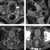
62-year-old male with muscle-invasive plasmacytoid urothelial carcinoma who underwent preoperative staging magnetic resonance imaging. (a) Axial and (b) sagittal T2-weighted turbo spin echoimages show residual bladder tumour (white arrow) with invasion of the seminal vesicles (arrowhead). There are ill-defined strands of low T2 signal intensity material within the pelvis, forming sheets of tissue, consistent with peritoneal spread of tumour (open arrows); (c) post-gadolinium axial T1W fat-suppressed gradient echo shows that these sheets of tissue demonstrate solid enhancement; (d) contrast-enhanced computed tomography performed earlier demonstrates corresponding appearance on computed tomography.
Fig. 4.
Fig. 4.
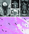
65-year-old male with plasmacytoid urothelial carcinoma (PUC) who underwent staging magnetic resonance imaging. Rectal examination at time of cystoscopy demonstrated pinpoint narrowing of the rectum, which was felt to be suspicious for synchronous rectal malignancy. (a) Axial T2-weighted turbo spin echo images show a heterogeneous low signal intensity (SI) mass invading through the posterior wall of the bladder into the seminal vesicles (white arrows). There are strands of low T2 SI in the pelvis (open arrows) with low T2 SI surrounding the rectum (arrowheads). Note that the normal rectal wall architecture is preserved; (b) axial b 1000 mm2/sec echo-planar images and axial apparent diffusion coefficient map show restricted diffusion in the bladder mass (white arrow) and in the rectal/perirectal soft tissues (arrowheads). Findings at pelvic exenteration confirmed locally advanced PUC. There was no rectal carcinoma; (c) contrast-enhanced computed tomography performed earlier demonstrates a similar pattern of spread, on retrospective review; (d) pathological image in a separate patient with PUC who underwent cystectomy, demonstrating typical pattern of spread with sheets of PUC cells (black arrows) invading the plane between the detrusor muscle (asterisk) and perivesical adipose tissue (black arrowheads).
Fig 5.
Fig 5.
75-year-old male with biopsy proven plasmacytoid urothelial carcinoma. (a) Axial contrast-enhanced computed tomography image through the upper abdomen obtained during the urographic phase of enhancement (split-bolus protocol) demonstrates large volume of malignant ascites with peritoneal carcinomatosis (arrows); (b) axial contrast-enhanced computed tomography image in the pelvis demonstrates diffuse bladder wall thickening with ill-defined soft tissue spreading into the pelvis with invasion of the seminal vesicles and prostate (arrows).

References

    1. Guzzo TJ, Malkowicz SB, Vaughn DJ, et al. Penn clinical manual of urology. 2nd edition. Philadelphia, PA: Saunders; 2014. Chapter 16 Adult genitourinary cancer; pp. 505–8.
    1. Wood DP. Campbell-Walsh Urology. 10th edition. Philadelphia, PA: Saunders; 2012. Chapter 80 Urothelial tumours of the bladder; pp. 2309–34.
    1. Lopez-Beltran A, Sauter G, Gasser T, et al. World health organization classification of tumours Pathology and genetics of tumours of the urinary system and male genital organs. Lyon: IARC Press; 2004. Infiltrating urothelial carcinoma; pp. 93–109.
    1. Sahin AA, Myhre M, Ro JY, et al. Plasmacytoid transitional cell carcinoma. Report of a case with initial presentation mimicking multiple myeloma. Acta Cytol. 1991;35:277–80. - PubMed
    1. Zukerberg LR, Harris NL, Young RH. Carcinomas of the urinary bladder simulating malignant lymphoma. A report of five cases. Am J Surg Pathol. 1991;15:569–76. https:/doi.org/10.1097/00000478-199106000-00005. - DOI - PubMed

LinkOut - more resources