

Proximal ureteral atresia, a rare congenital anomaly-incidental finding: a case report
- PMID: 28164035
- PMCID: PMC5253269
- DOI: 10.21037/tp.2017.01.02
Proximal ureteral atresia, a rare congenital anomaly-incidental finding: a case report
Abstract
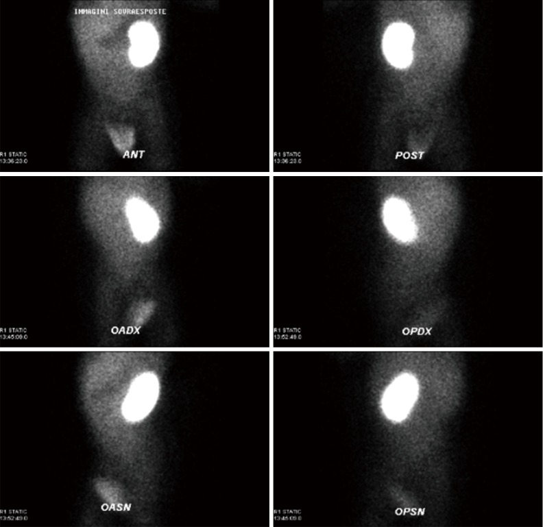
Ureteral atresia is a rare disease usually associated with a non-functioning dysplastic kidney. The condition may be unilateral or bilateral; focal, short or long and may involve any part of the ureter. Association with other urinary anomalies is rare. We report the case of a 10-month-old boy with prenatal diagnosis of multicystic right kidney. This suspicion was confirmed after birth by ultrasound and static scintigraphy; a right vesicoureteral reflux (VUR) was recorded at cystography. The boy presented a regular renal function but was hospitalized twice for suspected pyelonephritis between the 8th and the 10th month of life and were recorded occasional mild changes in blood pressure. Antibiotic prophylaxis was administered until surgery. When he was 10-month underwent retroperitoneoscopy to perform a nephroureterectomy finding a complete atresia of the upper third of the ureter with the blind end at the level of the uretero-pelvic-junction. The programmed surgery was performed. By a revision of literature, only few cases of imperforate distal ureter have been described. This condition is associated with a kidney dysplasia. The atresia of the ureter with no signs of infection in the dysplastic kidney may be unknown up to adulthood or throughout one's life. Prognosis usually depends on the severity of the obstruction.
Keywords: Dysplastic kidney; retroperitoneoscopy; ureteral atresia; ureteropelvic junction.
Conflict of interest statement
The authors have no conflicts of interest to declare.
Figures


References
-
- Sinha RS, Bhattacharjee P, Majhi T. Distal ureteric atresia—A Case Report. J Indian Assoc Pediatr Surg 2002;7:156-8.
Publication types
LinkOut - more resources
Full Text Sources
Other Literature Sources