. 2017 Jan 20:7:8.
doi: 10.3389/fcimb.2017.00008. eCollection 2017.

TLR2-/- Mice Display Increased Clearance of Dermatophyte Trichophyton mentagrophytes in the Setting of Hyperglycemia

Affiliations

TLR2-/- Mice Display Increased Clearance of Dermatophyte Trichophyton mentagrophytes in the Setting of Hyperglycemia

Débora de Fátima Almeida et al. Front Cell Infect Microbiol. .

Abstract

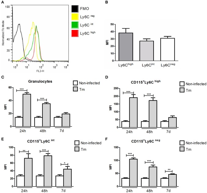
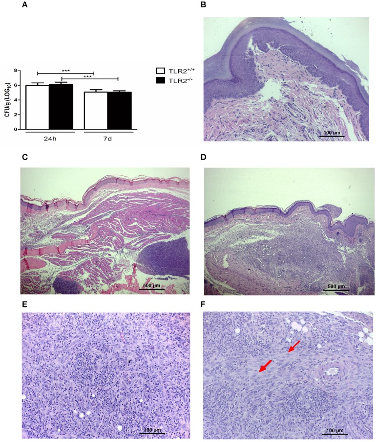
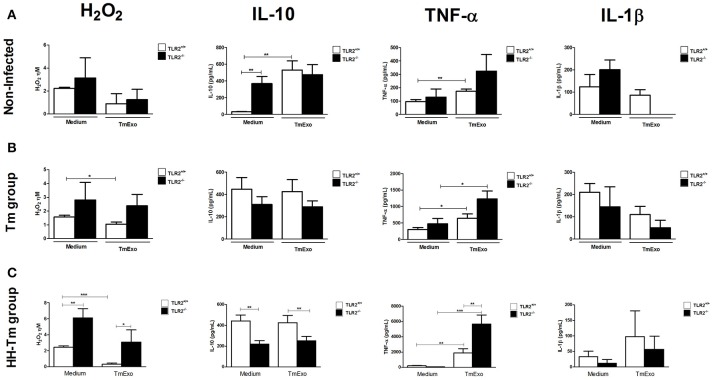
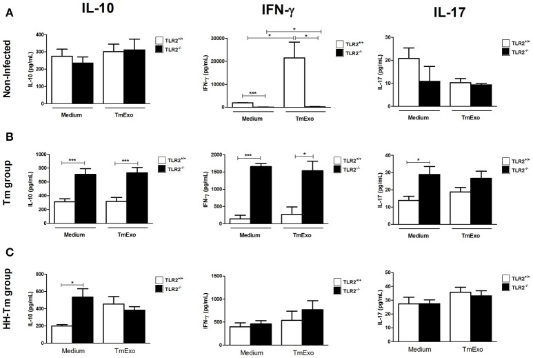
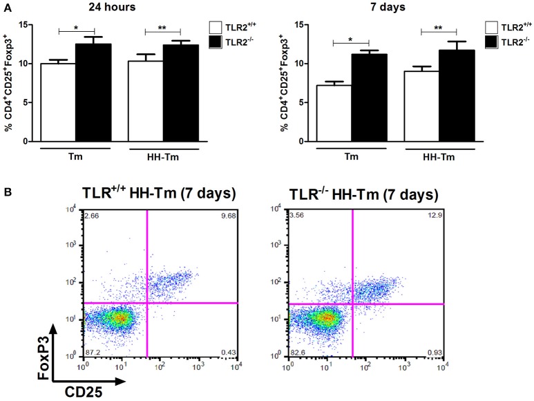
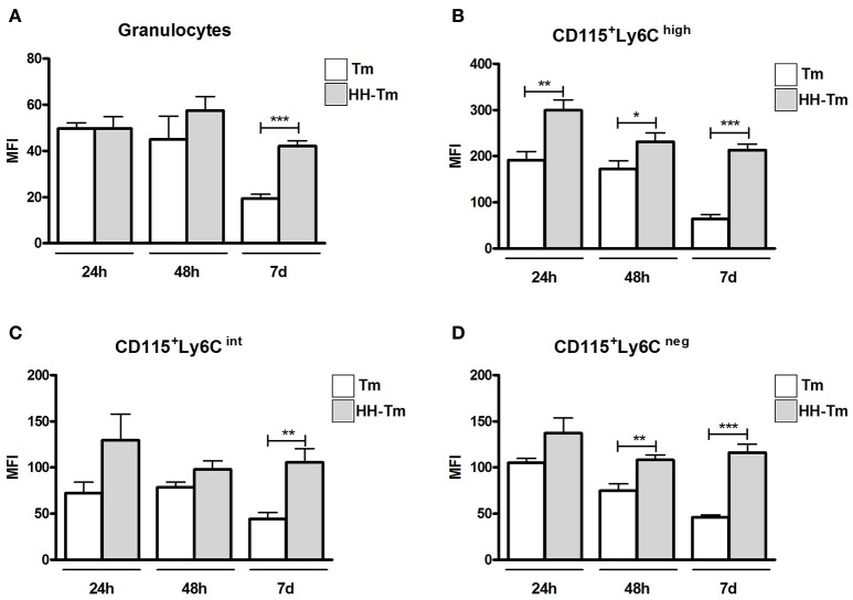
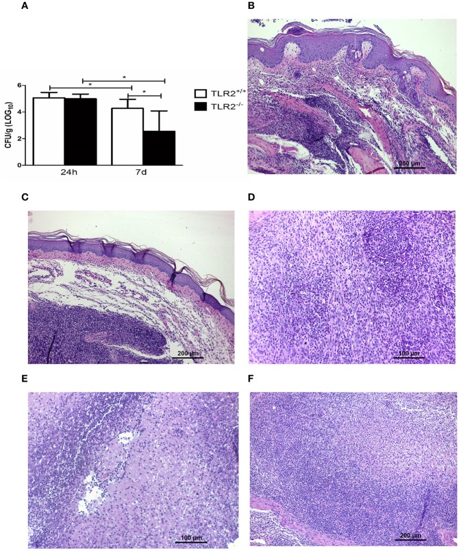
Dermatophytosis is one of the most common human infections affecting both immunocompetent individuals and immunocompromised patients, in whom the disease is more aggressive and can reach deep tissues. Over the last decades, cases of deep dermatophytosis have increased and the dermatophyte-host interplay remains poorly investigated. Pattern recognition molecules, such as Toll-like receptors (TLR), play a crucial role against infectious diseases. However, there has been very little research reported on dermatophytosis. In the present study, we investigated the role of TLR2 during the development of experimental deep dermatophytosis in normal mice and mice with alloxan-induced diabetes mellitus, an experimental model of diabetes that exhibits a delay in the clearance of the dermatophyte, Trichophyton mentagrophytes (Tm). Our results demonstrated that inoculation of Tm into the footpads of normal mice increases the expression of TLR2 in CD115+Ly6Chigh blood monocytes and, in hypoinsulinemic-hyperglycemic (HH) mice infected with Tm, the increased expression of TLR2 was exacerbated. To understand the role of TLR2 during the development of murine experimental deep dermatophytosis, we employed TLR2 knockout mice. Tm-infected TLR2-/- and TLR2+/+ wild-type mice exhibited similar control of deep dermatophytic infection and macrophage activity; however, TLR2-/- mice showed a noteworthy increase in production of IFN-γ, IL-10, and IL-17, and an increased percentage of splenic CD25+Foxp3+ Treg cells. Interestingly, TLR2-/- HH-Tm mice exhibited a lower fungal load and superior organization of tissue inflammatory responses, with high levels of production of hydrogen peroxide by macrophages, alongside low TNF-α and IL-10; high production of IL-10 by spleen cells; and increased expansion of Tregs. In conclusion, we demonstrate that TLR2 diminishes the development of adaptive immune responses during experimental deep dermatophytosis and, in a diabetic scenario, acts to intensify a non-protective inflammatory response.

Keywords: Trichophyton mentagrophytes; deep dermatophytosis; hypoinsulinemia-hyperglycemia; monocytes; toll-like receptors.


Figures

Figure 1
Figure 1
Expression of TLR2 in peripheral blood granulocytes and monocyte subsets from Swiss mice. Histogram (A) and mean fluorescence intensity (B) of TLR2 expression in monocyte subsets of naïve mice. Expression of TLR2 in granulocytes (C) and monocyte subsets (D–F) of non-infected and Tm-infected mice, evaluated at 24 and 48 h, and 7 days after infection. The results are expressed as means ± SEM (Unpaired t-test; *P < 0.05, **P < 0.01, ***P < 0.001; n = 5–6).
Figure 2
Figure 2
The dynamics of fungal infection in Tm-infected TLR2+/+ and TLR2−/− mice. (A) Recovery of viable fungi from footpads of Tm-infected mice 24 h and 7 days p.i. Results are expressed as mean ± SD (Unpaired t-test: ***P < 0.001). (B) Footpad of a naïve mouse (HE). (C) Footpad of a TLR2+/+ mouse 24 h after inoculation, showing a typical acute inflammatory response, characterized by an influx of neutrophils, edema, and abscess formation (HE). (D) Footpad of a TLR2+/+ mouse 7 days after infection, showing an initial granulomatous reaction with a small number of neutrophils and modified macrophages; details are shown in (E) (HE). (F) Footpad of a TLR2−/− mouse 7 days after infection, showing a well-organized granulomatous response, characterized by modified macrophages and epithelioid cells (arrow) (HE).
Figure 3
Figure 3
Macrophage activity in naïve, Tm-infected, and HH-Tm TLR2+/+ and TLR2−/− mice. Peritoneal adherent cells were cultured in the presence or absence of TmExo and, after 24 h, the levels of hydrogen peroxide (H2O2), TNF-α, and IL-1β were determined in cell-free supernatants. (A) TLR2+/+ and TLR2−/− naïve mice, (B) Tm-infected TLR2+/+ and TLR2−/− mice, (C) HH-Tm TLR2+/+ and TLR2−/− mice. Results are expressed as means ± SEM. (Unpaired t-test; *P < 0.05, **P < 0.01, ***P < 0.001; n = 5–6).
Figure 4
Figure 4
Activity of splenic cells from naïve, Tm-infected, and HH-Tm TLR2+/+ and TLR2−/− mice. Spleen cells collected 7 days p.i. were cultured in the presence or absence of TmExo and the levels of IL-10, IFN-γ, and IL-17 were determined in the cell-free supernatants. (A) TLR2+/+ and TLR2−/− naïve mice, (B) Tm-infected TLR2+/+ and TLR2−/− mice, (C) HH-Tm TLR2+/+ and TLR2−/− mice. Results are expressed as means ± SEM (Unpaired t-test; *P < 0.05, ***P < 0.001; n = 5–6).
Figure 5
Figure 5
The distribution of CD25+Foxp3+ regulatory T lymphocytes in spleen cells. (A) Tm and HH-Tm TLR2+/+ and TLR2−/− mice were evaluated 24 h and 7 days after infection. Results are expressed as means ± SEM (Unpaired t-test: *P < 0.05, ** 0.01 < P < 0.05). (B) Representative dot plots of the gating strategy for CD25+Foxp3+.
Figure 6
Figure 6
Expression of TLR2 in peripheral blood granulocytes and monocyte subsets from Swiss mice. (A) Granulocytes and monocyte subsets (B–D) of Tm-infected and HH-Tm mice evaluated at 24 and 48 h, and 7 days, after infection. The results are expressed as means ± SEM (Unpaired t-test; *P < 0.05, **P < 0.01, ***P < 0.001; n = 5–6).
Figure 7
Figure 7
The dynamics of fungal infection in HH-Tm TLR2+/+ and TLR2−/− mice. (A) Recovery of viable fungi from footpads of HH-Tm mice 24 h and 7 days p.i. Results are expressed as means ± SD. Unpaired t-test: *P < 0.05. (B) Footpad of a TLR2+/+ HH infected mouse 24 h p.i. exhibiting the initial inflammatory pattern characterized by intense polymorphonuclear infiltrate and edema (HE). (C) Footpad of a TLR2−/− HH infected mouse 24 h p.i. exhibiting the initial inflammatory pattern characterized by intense polymorphonuclear infiltrate and edema (HE). (D) Footpad of a TLR2+/+ HH infected mouse on day 7 p.i. showing a mixed inflammatory infiltrate with a small number of differentiated macrophages and an intense influx of neutrophils (HE). (E) Footpad of a TLR2+/+ HH infected mouse showing abscess formation and necrosis (HE). (F) Footpad of a TLR2−/− HH infected mouse on day 7 p.i., showing more organized granulomas, with a small number of neutrophils, and the presence of modified macrophages (HE).

Similar articles

Cited by

References

    1. Abd elaziz E. A. (2011). Pathological and biochemical studies on the effect of trigonella foenum -graecum and lupinus termis in alloxan induced diabetic rats. World Appl. Sci. J. 12, 1839–1850.
    1. Almeida S. R. (2008). Immunology of dermatophytosis. Mycopathologia 166, 277–283. 10.1007/s11046-008-9103-6 - DOI - PubMed
    1. Calderon R. A., Hay R. J. (1984). Cell-mediated immunity in experimental murine dermatophytosis. II. Adoptive transfer of immunity to dermatophyte infection by lymphoid cells from donors with acute or chronic infections. Immunology 53, 465–472. - PMC - PubMed
    1. Campos M. R., Russo M., Gomes E., Almeida S. R. (2006). Stimulation, inhibition and death of macrophages infected with Trichophyton rubrum. Microbes Infect. 8, 372–379. 10.1016/j.micinf.2005.07.028 - DOI - PubMed
    1. Devaraj S., Tobias P., Kasinath B. S., Ramsamooj R., Afify A., Jialal I. (2011). Knockout of toll-like receptor-2 attenuates both the proinflammatory state of diabetes and incipient diabetic nephropathy. Arterioscler. Thromb. Vasc. Biol. 31, 1796–1804. 10.1161/ATVBAHA.111.228924 - DOI - PMC - PubMed