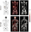
Multimodality molecular imaging of the lung
- PMID: 28164069
- PMCID: PMC5289702
- DOI: 10.1007/s40336-014-0084-9
Multimodality molecular imaging of the lung
Abstract
Lung diseases cause significant morbidity and mortality and lead to high healthcare utilization. However, few lung disease-specific biomarkers are available to accurately monitor disease activity for the purposes of clinical management or drug development. Advances in cross-modal imaging technologies, such as combined positron emission tomography (PET) and magnetic resonance (MR) imaging scanners and PET or single-photon emission computed tomography (SPECT) combined with computed tomography (CT), may aid in the development of noninvasive, molecular-based biomarkers for lung disease. However, the lungs pose particular challenges in obtaining accurate quantification of imaging data due to the low density of the organ and breathing motion. This review covers the basic physics underlying PET, SPECT, CT, and MR lung imaging and presents technical considerations for multimodal imaging with regard to PET and SPECT quantification. It also includes a brief review of the current and potential clinical applications for these hybrid imaging technologies.
Keywords: Lung cancer; Lung disease; Magnetic resonance imaging; Molecular imaging; Multimodality imaging; Positron emission tomography; Single photon emission computed tomography.
Conflict of interest statement
Neither Delphine L. Chen nor Paul E. Kinahan have any conflicts of interest to declare.
Figures


References
-
- National Heart Lung Blood Institute. [Accessed 26 Sept 2014];Morbidity and Mortality: 2012 Chart Book on Cardiovascular, lung, and blood diseases. 2012 https://www.nhlbi.nih.gov/files/docs/research/2012_ChartBook.pdf.
-
- American Cancer Society. [Accessed 26 Sept 2014];Cancer facts and figures 2014. 2014 http://www.cancer.org/acs/groups/content/@research/documents/webcontent/....
-
- Martinez FJ, Donohue JF, Rennard SI. The future of chronic obstructive pulmonary disease treatment—difficulties of and barriers to drug development. Lancet. 2011;37:1027–1037. - PubMed
-
- Cherry SR, Sorenson JA, Phelps ME. Physics in nuclear medicine. 4. Saunders; Philadelphia: 2012.
Grants and funding
LinkOut - more resources
Full Text Sources
Other Literature Sources
