Review
. 2017 Jan;5(1):3.
doi: 10.21037/atm.2016.12.78.

Effective implementation of novel MET pharmacodynamic assays in translational studies

Affiliations
Review

Effective implementation of novel MET pharmacodynamic assays in translational studies

Apurva K Srivastava et al. Ann Transl Med. 2017 Jan.

Abstract

MET tyrosine kinase (TK) dysregulation is significantly implicated in many types of cancer. Despite over 20 years of drug development to target MET in cancers, a pure anti-MET therapeutic has not yet received market approval. The failure of two recently concluded phase III trials point to a major weakness in biomarker strategies to identify patients who will benefit most from MET therapies. The capability to interrogate oncogenic mutations in MET via circulating tumor DNA (ctDNA) provides an important advancement in identification and stratification of patients for MET therapy. However, a wide range in type and frequency of these mutations suggest there is a need to carefully link these mutations to MET dysregulation, at least in proof-of-concept studies. In this review, we elaborate how we can utilize recently developed and validated pharmacodynamic biomarkers of MET not only to show target engagement, but more importantly to quantitatively measure MET dysregulation in tumor tissues. The MET assay endpoints provide evidence of both canonical and non-canonical MET signaling, can be used as "effect markers" to define biologically effective doses (BEDs) for molecularly targeted drugs, confirm mechanism-of-action in testing combination of drugs, and establish whether a diagnostic test is reporting MET dysregulation. We have established standard operating procedures for tumor biopsy collections to control pre-analytical variables that have produced valid results in proof-of-concept studies. The reagents and procedures are made available to the research community for potential implementation on multiple platforms such as ELISA, quantitative immunofluorescence assay (qIFA), and immuno-MRM assays.

Keywords: MET; immunoassay; pharmacodynamic assay; phosphorylation; preclinical model; translational biomarker.


Conflict of interest statement

The authors have no conflicts of interest to declare.

Figures
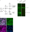
Figure 1
Figure 1
Phosphorylated A-loop tyrosine Y1235 is preferentially phosphorylated in some mutant forms of MET and appears to be processed differentially. (A) In the WT MET receptor, phosphorylation of the two A-loop tyrosines Y1234 and Y1235 is necessary to induce MET canonical signaling. In HPRC cells, oncogenic mutations in the MET gene can overcome the need for phosphorylation of Y1234, and kinase activity of MET can be achieved by phosphorylation of Y1235 only. We developed an antibody specific to pY1235 that binds MET irrespective of Y1234 phosphorylation. (B) Using this antibody we demonstrated that the mono-phosphorylated pY1235-MET is processed differentially as a smaller fragment and (C) accumulates in the nucleus. WT, wild type; HPRC, hereditary papillary renal carcinoma. (Figure 1B,C reproduced from Srivastava et al., Clin Can Res, 2016).
Figure 2
Figure 2
Lung cancer model H596 tumor xenografts (harboring MET exon 14 skipping mutations) from hHGF knockin SCID mice exhibit enhanced nuclear pY1235-MET expression compared to tumors obtained from wild type SCID mice. (A) H596 cells (1×10⁷) were implanted in either SCID (Prkdcscid), hHGFki/− (human HGF knockin) or hHGFki/ki mice and tumor growth monitored 2–3 times per week for the indicated number of days. Tumors were harvested after they have reached 300 mg in tumor weight; (B) top row panel shows representative monochromatic IFA image of TMA tumor tissues derived from hHGF knockin and SCID mice showing nuclear pY1235-MET expression. Middle and lower panels show pY1235-MET (red) and nuclei (DAPI; blue) marker masks, respectively, derived from Definiens image analysis; (C) scatter graph shows significant increases in nuclear pY1235-MET, expressed as nuclear area percentage (%NAP), in H596 tumors obtained from HGF knockin vs. SCID mice. Each point corresponds to measurements obtained from an individual mouse. *, P<0.016 vs. SCID (not adjusted for multiple comparison); **, P<0.0095 vs. SCID; (D) high resolution image of focal nuclear pY1235-MET IFA staining in the nuclei of H596 tumor xenografts; (E) representative immunofluorescence image of H596 tumor tissues derived from SCID and hHGF knockin mice stained by IFA for E-cadherin (green) and vimentin (red). The images show increased mesenchymal tumor phenotype in the HGF knockin mice compared to the SCID background.
Figure 3
Figure 3
Magnitude of phosphorylation at Y1234 and Y1235 in MET amplified cell line SNU5 and tumor biopsies. (A) In the MET amplified SNU5 cell line, the magnitude of phosphorylation of two A-loop tyrosines Y1234 and Y1235 as measured by two separate ELISA. A pY1235-MET specific ELISA (clone 23111) measured only Y1235-MET phosphorylation, whereas, pY1234/1235-MET ELISA detected both pY1234-MET and pY1235-MET. The percentage inhibition of both tyrosines (clone D26) by a MET inhibitor, crizotinib, was comparable; (B) the differential phosphorylation ratio of A-loop tyrosine was confirmed in an MRM mass spectrometric assay employing isotope labeled peptide calibrators. Top panel shows mass peaks from cell lysates and bottom panel shows corresponding peaks from heavy peptide calibrators; (C) ratio of full length dual pY1234/35-MET/MET in biopsies of a sporadic renal tumor from HPRC. RT, retention time; AA, peak area. (Figure 3A recreated from Srivastava et al., Clin Can Res, 2016).

References

    1. Gherardi E, Birchmeier W, Birchmeier C, et al. Targeting MET in cancer: rationale and progress. Nat Rev Cancer 2012;12:89-103. 10.1038/nrc3205 - DOI - PubMed
    1. Comoglio PM, Giordano S, Trusolino L. Drug development of MET inhibitors: targeting oncogene addiction and expedience. Nat Rev Drug Discov 2008;7:504-16. 10.1038/nrd2530 - DOI - PubMed
    1. Rolfo C, Van Der Steen N, Pauwels P, et al. Onartuzumab in lung cancer: the fall of Icarus? Expert Rev Anticancer Ther 2015;15:487-9. 10.1586/14737140.2015.1031219 - DOI - PubMed
    1. Scagliotti G, von Pawel J, Novello S, et al. Phase III Multinational, Randomized, Double-Blind, Placebo-Controlled Study of Tivantinib (ARQ 197) Plus Erlotinib Versus Erlotinib Alone in Previously Treated Patients With Locally Advanced or Metastatic Nonsquamous Non-Small-Cell Lung Cancer. J Clin Oncol 2015;33:2667-74. 10.1200/JCO.2014.60.7317 - DOI - PubMed
    1. Yoshioka H, Azuma K, Yamamoto N, et al. A randomized, double-blind, placebo-controlled, phase III trial of erlotinib with or without a c-Met inhibitor tivantinib (ARQ 197) in Asian patients with previously treated stage IIIB/IV nonsquamous nonsmall-cell lung cancer harboring wild-type epidermal growth factor receptor (ATTENTION study). Ann Oncol 2015;26:2066-72. 10.1093/annonc/mdv288 - DOI - PubMed

LinkOut - more resources