

Plumbagin Suppresses α-MSH-Induced Melanogenesis in B16F10 Mouse Melanoma Cells by Inhibiting Tyrosinase Activity
- PMID: 28165370
- PMCID: PMC5343856
- DOI: 10.3390/ijms18020320
Plumbagin Suppresses α-MSH-Induced Melanogenesis in B16F10 Mouse Melanoma Cells by Inhibiting Tyrosinase Activity
Abstract
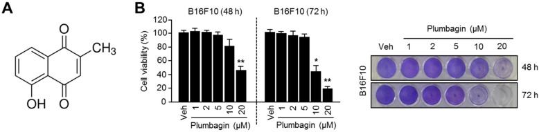
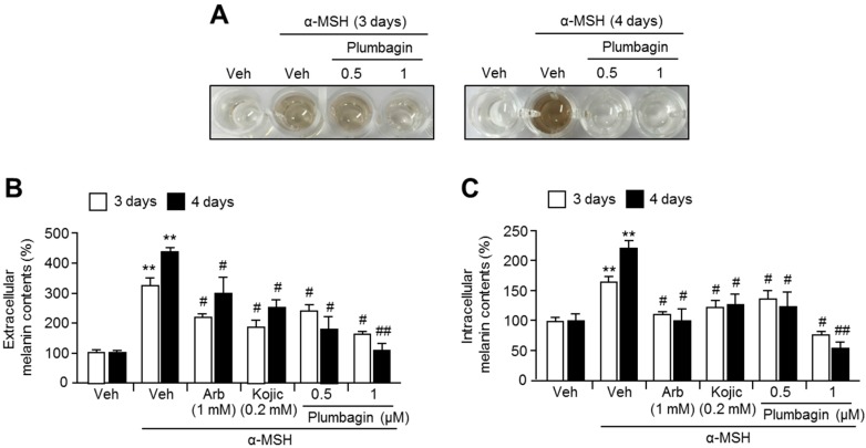
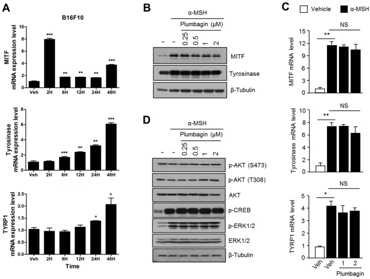
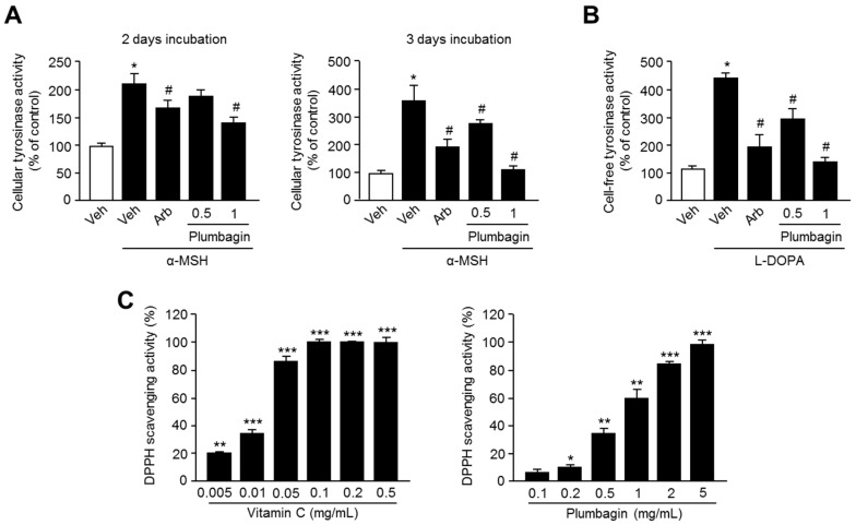
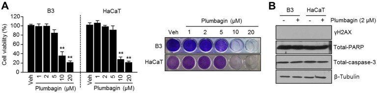
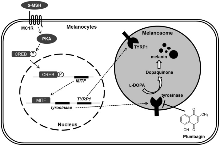
Recent studies have shown that plumbagin has anti-inflammatory, anti-allergic, antibacterial, and anti-cancer activities; however, it has not yet been shown whether plumbagin suppresses alpha-melanocyte stimulating hormone (α-MSH)-induced melanin synthesis to prevent hyperpigmentation. In this study, we demonstrated that plumbagin significantly suppresses α-MSH-stimulated melanin synthesis in B16F10 mouse melanoma cells. To understand the inhibitory mechanism of plumbagin on melanin synthesis, we performed cellular or cell-free tyrosinase activity assays and analyzed melanogenesis-related gene expression. We demonstrated that plumbagin directly suppresses tyrosinase activity independent of the transcriptional machinery associated with melanogenesis, which includes micropthalmia-associated transcription factor (MITF), tyrosinase (TYR), and tyrosinase-related protein 1 (TYRP1). We also investigated whether plumbagin was toxic to normal human keratinocytes (HaCaT) and lens epithelial cells (B3) that may be injured by using skin-care cosmetics. Surprisingly, lower plumbagin concentrations (0.5-1 μM) effectively inhibited melanin synthesis and tyrosinase activity but do not cause toxicity in keratinocytes, lens epithelial cells, and B16F10 mouse melanoma cells, suggesting that plumbagin is safe for dermal application. Taken together, these results suggest that the inhibitory effect of plumbagin to pigmentation may make it an acceptable and safe component for use in skin-care cosmetic formulations used for skin whitening.
Keywords: melanogenesis; pigmentation; plumbagin; tyrosinase.
Conflict of interest statement
The authors declare no conflict of interest.
Figures
Similar articles
-
Curcumin suppresses alpha-melanocyte stimulating hormone-stimulated melanogenesis in B16F10 cells.Int J Mol Med. 2010 Jul;26(1):101-6. Int J Mol Med. 2010. PMID: 20514428
-
Acteoside inhibits melanogenesis in B16F10 cells through ERK activation and tyrosinase down-regulation.J Pharm Pharmacol. 2011 Oct;63(10):1309-19. doi: 10.1111/j.2042-7158.2011.01335.x. Epub 2011 Aug 19. J Pharm Pharmacol. 2011. PMID: 21899547
-
Inhibition of tyrosinase activity and melanin production by the chalcone derivative 1-(2-cyclohexylmethoxy-6-hydroxy-phenyl)-3-(4-hydroxymethyl-phenyl)-propenone.Biochem Biophys Res Commun. 2016 Nov 25;480(4):648-654. doi: 10.1016/j.bbrc.2016.10.110. Epub 2016 Oct 27. Biochem Biophys Res Commun. 2016. PMID: 27983977
-
Recent development of signaling pathways inhibitors of melanogenesis.Cell Signal. 2017 Dec;40:99-115. doi: 10.1016/j.cellsig.2017.09.004. Epub 2017 Sep 12. Cell Signal. 2017. PMID: 28911859 Review.
-
Natural tyrosinase enzyme inhibitors: A path from melanin to melanoma and its reported pharmacological activities.Biochim Biophys Acta Rev Cancer. 2023 Nov;1878(6):188968. doi: 10.1016/j.bbcan.2023.188968. Epub 2023 Aug 30. Biochim Biophys Acta Rev Cancer. 2023. PMID: 37657683 Review.
Cited by
-
Ribosomal S6 kinase 2-forkhead box protein O4 signaling pathway plays an essential role in melanogenesis.Sci Rep. 2024 Apr 24;14(1):9440. doi: 10.1038/s41598-024-60165-9. Sci Rep. 2024. PMID: 38658799 Free PMC article.
-
Antimelanogenic Effects of Polygonum tinctorium Flower Extract from Traditional Jeju Fermentation via Upregulation of Extracellular Signal-Regulated Kinase and Protein Kinase B Activation.Int J Mol Sci. 2018 Sep 24;19(10):2895. doi: 10.3390/ijms19102895. Int J Mol Sci. 2018. PMID: 30249988 Free PMC article.
-
Design, Synthesis, and Anti-Tyrosinase, Anti-Melanogenic, and Antioxidant Activities of Novel (Z)-3-Benzyl-5-Benzylidene-2-Thioxothiazolidin-4-One Analogs.Molecules. 2025 Jan 23;30(3):517. doi: 10.3390/molecules30030517. Molecules. 2025. PMID: 39942621 Free PMC article.
-
Low Molecular Weight Oligosaccharide from Panax ginseng C.A. Meyer against UV-Mediated Apoptosis and Inhibits Tyrosinase Activity In Vitro and In Vivo.Evid Based Complement Alternat Med. 2021 Feb 26;2021:8879836. doi: 10.1155/2021/8879836. eCollection 2021. Evid Based Complement Alternat Med. 2021. PMID: 33727947 Free PMC article.
-
Anti-Melanogenic Effects of a Polysaccharide Isolated from Undaria pinnatifida Sporophyll Extracts.Int J Mol Sci. 2024 Oct 2;25(19):10624. doi: 10.3390/ijms251910624. Int J Mol Sci. 2024. PMID: 39408953 Free PMC article.
References
-
- Kang S.J., Choi B.R., Lee E.K., Kim S.H., Yi H.Y., Park H.R., Song C.H., Lee Y.J., Ku S.K. Inhibitory effect of dried pomegranate concentration powder on melanogenesis in B16F10 melanoma cells; involvement of p38 and PKA signaling pathways. Int. J. Mol. Sci. 2015;16:24219–24242. doi: 10.3390/ijms161024219. - DOI - PMC - PubMed
MeSH terms
Substances
LinkOut - more resources
Full Text Sources
Other Literature Sources
Research Materials
