

Bone Mineral Density, Mechanical, Microstructural Properties and Mineral Content of the Femur in Growing Rats Fed with Cactus Opuntia ficus indica (L.) Mill. (Cactaceae) Cladodes as Calcium Source in Diet
- PMID: 28165410
- PMCID: PMC5331539
- DOI: 10.3390/nu9020108
Bone Mineral Density, Mechanical, Microstructural Properties and Mineral Content of the Femur in Growing Rats Fed with Cactus Opuntia ficus indica (L.) Mill. (Cactaceae) Cladodes as Calcium Source in Diet
Abstract
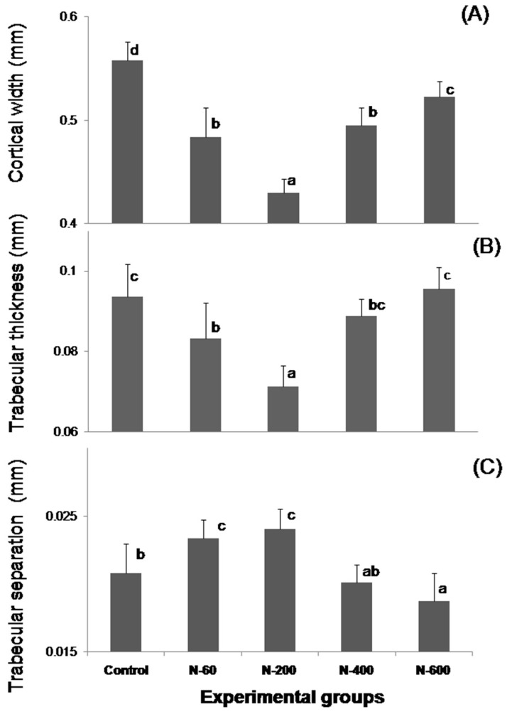
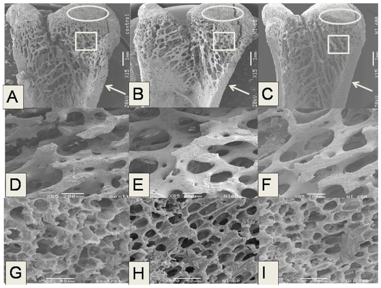
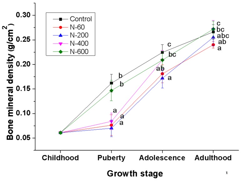
Mechanical, microstructural properties, mineral content and bone mineral density (BMD) of the femur were evaluated in growing rats fed with Opuntia ficus indica (L.) Mill. (Cactaceae) cladodes at different maturity stages as calcium source. Male weanling rats were fed with cladodes at early maturity stage (25 and 60 days of age, belonging to groups N-60 and N-200, respectively) and cladodes at late maturity stage (100 and 135 days of age, belonging to groups N-400 and N-600, respectively) for 6 weeks. Additionally, a control group fed with calcium carbonate as calcium source was included for comparative purposes. All diets were fitted to the same calcium content (5 g/kg diet). The failure load of femurs was significantly lower (p ≤ 0.05) in groups N-60 and N-200 in comparison to N-400, N-600 and control groups. The cortical width (Ct.Wi) and trabecular thickness (Tb.Th) of the femurs in control and N-600 groups were significantly higher (p ≤ 0.05) than Ct.Wi and Tb.Th of femurs in groups N-60 and N-200. Trabecular separation of the femurs in N-60 and N-200 groups showed the highest values compared with all experimental groups. The highest calcium content in the femurs were observed in control, N-600 and N-400 groups; whereas the lowest phosphorus content in the bones were detected in N-200, N-600 and N-400 groups. Finally, the BMD in all experimental groups increased with age; nevertheless, the highest values were observed in N-600 and control groups during pubertal and adolescence stages. The results derived from this research demonstrate, for the first time, that the calcium found in Opuntia ficus indica cladodes is actually bioavailable and capable of improving mineral density and mechanical and microstructural properties of the bones. These findings suggest that the consumption of cladodes at late maturity stage within the diet might have a beneficial impact on bone health.
Keywords: Opuntia ficus indica; bone mineral density; cactus; calcium; maturity stage; microstructure; phosphorus.
Conflict of interest statement
The authors declare no conflict of interest.
Figures
Similar articles
-
Calcium Bioavailability in the Soluble and Insoluble Fibers Extracted from Opuntia ficus indica at Different Maturity Stages in Growing Rats.Nutrients. 2020 Oct 23;12(11):3250. doi: 10.3390/nu12113250. Nutrients. 2020. PMID: 33114068 Free PMC article.
-
Calcium Bioavailability of Opuntia ficus-indica Cladodes in an Ovariectomized Rat Model of Postmenopausal Bone Loss.Nutrients. 2020 May 15;12(5):1431. doi: 10.3390/nu12051431. Nutrients. 2020. PMID: 32429103 Free PMC article.
-
Effect of Nopal (Opuntia ficus indica) Consumption at Different Maturity Stages as an Only Calcium Source on Bone Mineral Metabolism in Growing Rats.Biol Trace Elem Res. 2020 Mar;194(1):168-176. doi: 10.1007/s12011-019-01752-0. Epub 2019 May 24. Biol Trace Elem Res. 2020. PMID: 31127473
-
Opuntia ficus-indica (L.) Mill. - anticancer properties and phytochemicals: current trends and future perspectives.Front Plant Sci. 2023 Oct 4;14:1236123. doi: 10.3389/fpls.2023.1236123. eCollection 2023. Front Plant Sci. 2023. PMID: 37860248 Free PMC article. Review.
-
Opuntia ficus-indica (L.) Miller as a source of bioactivity compounds for health and nutrition.Nat Prod Res. 2018 Sep;32(17):2037-2049. doi: 10.1080/14786419.2017.1365073. Epub 2017 Aug 14. Nat Prod Res. 2018. PMID: 28805459 Review.
Cited by
-
Sex- and Age-Related Dynamic Changes of the Macroelements Content in the Femoral Bone with Hip Osteoarthritis.Biology (Basel). 2022 Feb 22;11(3):344. doi: 10.3390/biology11030344. Biology (Basel). 2022. PMID: 35336718 Free PMC article.
-
Calcium Bioavailability in the Soluble and Insoluble Fibers Extracted from Opuntia ficus indica at Different Maturity Stages in Growing Rats.Nutrients. 2020 Oct 23;12(11):3250. doi: 10.3390/nu12113250. Nutrients. 2020. PMID: 33114068 Free PMC article.
-
Comparative Analysis of the Chemical Composition and Physicochemical Properties of the Mucilage Extracted from Fresh and Dehydrated Opuntia ficus indica Cladodes.Foods. 2021 Sep 10;10(9):2137. doi: 10.3390/foods10092137. Foods. 2021. PMID: 34574247 Free PMC article.
-
Amorphous Calcium Carbonate from Plants Can Promote Bone Growth in Growing Rats.Biology (Basel). 2024 Mar 21;13(3):201. doi: 10.3390/biology13030201. Biology (Basel). 2024. PMID: 38534470 Free PMC article.
-
Calcium Bioavailability of Opuntia ficus-indica Cladodes in an Ovariectomized Rat Model of Postmenopausal Bone Loss.Nutrients. 2020 May 15;12(5):1431. doi: 10.3390/nu12051431. Nutrients. 2020. PMID: 32429103 Free PMC article.
References
MeSH terms
Substances
LinkOut - more resources
Full Text Sources
Other Literature Sources
Medical
