. 2017 Feb 6:2:16269.
doi: 10.1038/nmicrobiol.2016.269.

Architecture of the Vibrio cholerae toxin-coregulated pilus machine revealed by electron cryotomography

Affiliations

Architecture of the Vibrio cholerae toxin-coregulated pilus machine revealed by electron cryotomography

Yi-Wei Chang et al. Nat Microbiol. .

Abstract

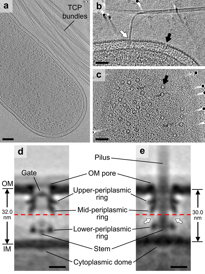
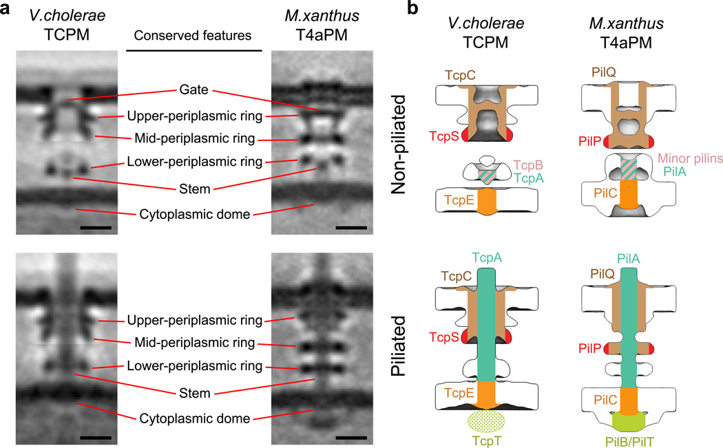
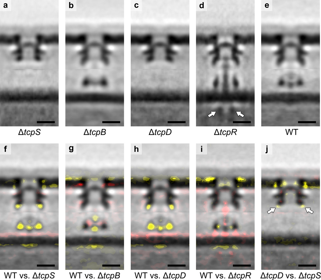
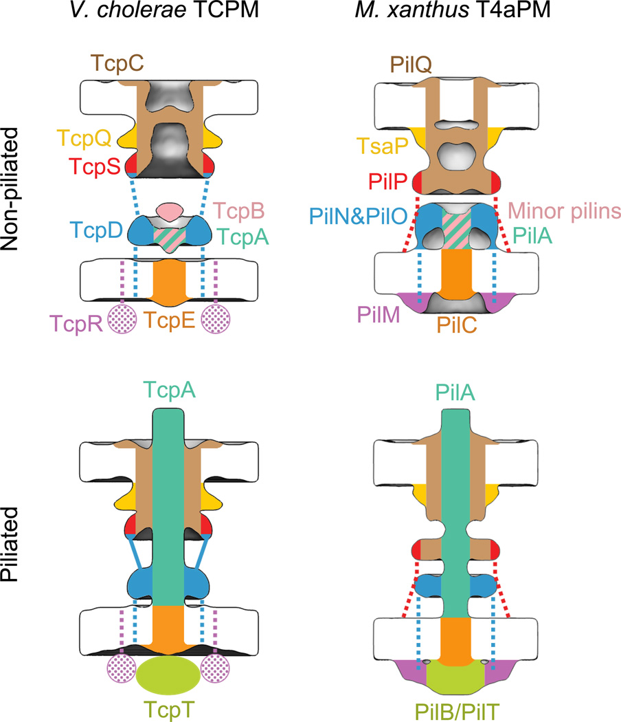
Type IV pili (T4P) are filamentous appendages found on many Bacteria and Archaea. They are helical fibres of pilin proteins assembled by a multi-component macromolecular machine we call the basal body. Based on pilin features, T4P are classified into type IVa pili (T4aP) and type IVb pili (T4bP)1,2. T4aP are more widespread and are involved in cell motility3, DNA transfer4, host predation5 and electron transfer6. T4bP are less prevalent and are mainly found in enteropathogenic bacteria, where they play key roles in host colonization7. Following similar work on T4aP machines8,9, here we use electron cryotomography10 to reveal the three-dimensional in situ structure of a T4bP machine in its piliated and non-piliated states. The specific machine we analyse is the Vibrio cholerae toxin-coregulated pilus machine (TCPM). Although only about half of the components of the TCPM show sequence homology to components of the previously analysed Myxococcus xanthus T4aP machine (T4aPM), we find that their structures are nevertheless remarkably similar. Based on homologies with components of the M. xanthus T4aPM and additional reconstructions of TCPM mutants in which the non-homologous proteins are individually deleted, we propose locations for all eight TCPM components within the complex. Non-homologous proteins in the T4aPM and TCPM are found to form similar structures, suggesting new hypotheses for their functions and evolutionary histories.


Figures

Figure 1
Figure 1. Visualizing the TCPM in intact V. cholerae cells
(a) Slice through a cryotomogram of a frozen-hydrated V. cholerae cell on an EM grid adjacent to bundles of TCP. (b) A tomographic slice through the cell envelope. White arrow: a piliated TCPM basal body with its pilus fiber joining a TCP bundle; black arrow: a non-piliated TCPM basal body. (c) A tomographic slice through the cell surface showing top views of TCPM OM pores (black arrow). Images in (a–c) are representative of at least 50 examples. (d, e) Composite sub-tomogram averages of wild-type non-piliated (d) and piliated (e) TCPM basal bodies, with upper and lower halves (separated by red dashed lines) aligned based on the OM- or IM-associated parts, respectively. The generation of the composite sub-tomogram averages is demonstrated in Supplementary Figure 2. Arrows in (d): densities connecting the mid- and lower-periplasmic rings. Scale bars (a) 100 nm, (b, c) 50 nm, (d, e) 10 nm.
Figure 2
Figure 2. Comparison between V. cholerae TCPM and M. xanthus T4aPM structures, and the inferred TCPM component locations based on the T4aPM component map
(a) Left column: slices through the composite sub-tomogram averages of V. cholerae wild-type non-piliated and piliated TCPM structures. Right column: slices through sub-tomogram averages of M. xanthus ΔpilB non-piliated and wild-type piliated T4aPM structures. Scale bars, 10 nm. (b) Inferred TCPM component locations of TcpC, TcpS, TcpB, TcpA and TcpE (left column) based on their identified analogy to PilQ, PilP, minor pilins, PilA and PilC, respectively, in the reported T4aPM component map (right column).
Figure 3
Figure 3. Structures of TCPM in ΔtcpS, ΔtcpB, ΔtcpD and ΔtcpR cells
(a, b, c, d, e) Central slices of sub-tomogram averages of TCPM basal bodies from ΔtcpS, ΔtcpB, ΔtcpD, ΔtcpR and wild-type cells, respectively. Arrows in (d) indicate the cytoplasmic disc-like density. (f, g, h, i) Differences in the ΔtcpS, ΔtcpB, ΔtcpD and ΔtcpR structures compared to the wild-type, respectively (addition and omission of densities are shown by red or yellow color, respectively, with weak to strong intensities corresponding to density differences from one to three standard deviations, overlaid on the wild-type sub-tomogram average). (j) Differences in the ΔtcpS structure compared to the ΔtcpD structure (difference maps overlaid on the ΔtcpD sub-tomogram average). Arrows indicate a larger decrease of the mid-periplasmic ring density caused by ΔtcpS than by ΔtcpD. Scale bars, 10 nm.
Figure 4
Figure 4. Locations of TCPM components and the comparison with M. xanthus T4aPM
Left column: schematics showing the component locations assigned for the piliated and non-piliated V. cholerae TCPM (this study). Right column: schematics showing the component locations identified in the piliated and non-piliated M. xanthus T4aPM. Dashed lines indicate known connections without visible electron density in sub-tomogram averages. Dotted colors indicate proposed protein locations.

References

    1. Strom MS, Lory S. Structure-function and biogenesis of the type IV pili. Annu Rev Microbiol. 1993;47:565–596. - PubMed
    1. Craig L, Pique ME, Tainer JA. Type IV pilus structure and bacterial pathogenicity. Nat Rev Microbiol. 2004;2:363–378. - PubMed
    1. Mattick JS. Type IV pili and twitching motility. Annu Rev Microbiol. 2002;56:289–314. - PubMed
    1. Chen I, Dubnau D. DNA uptake during bacterial transformation. Nat Rev Microbiol. 2004;2:241–249. - PubMed
    1. Evans KJ, Lambert C, Sockett RE. Predation by Bdellovibrio bacteriovorus HD100 requires type IV pili. J Bacteriol. 2007;189:4850–4859. - PMC - PubMed

MeSH terms