. 2017 Feb 6:8:14388.
doi: 10.1038/ncomms14388.

Structure of the homodimeric androgen receptor ligand-binding domain

Affiliations

Structure of the homodimeric androgen receptor ligand-binding domain

Marta Nadal et al. Nat Commun. .

Abstract

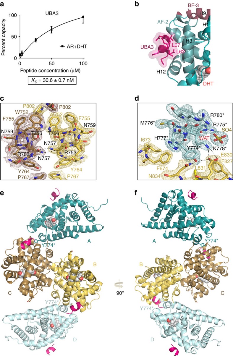
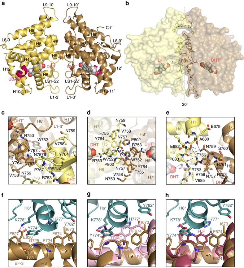
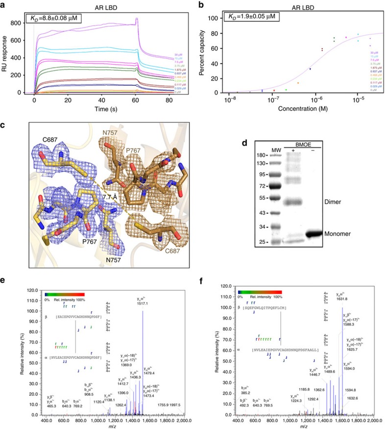
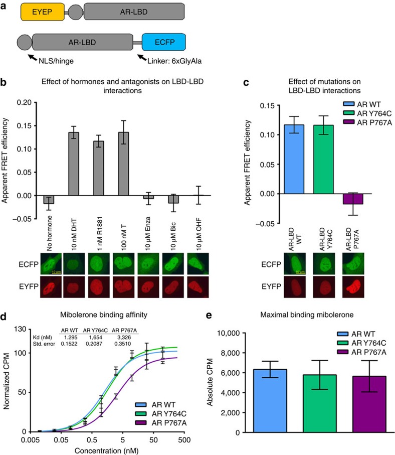
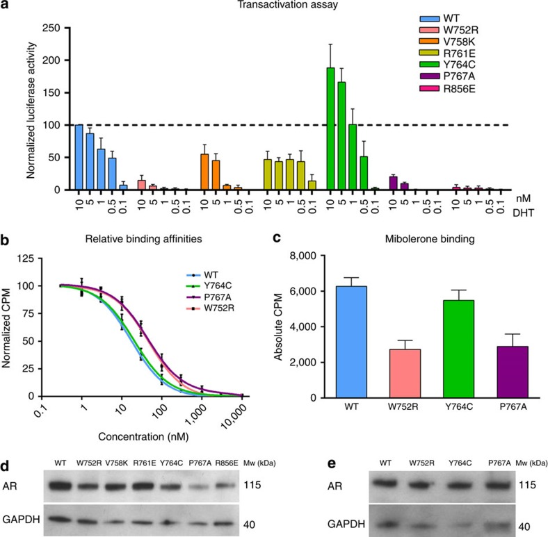
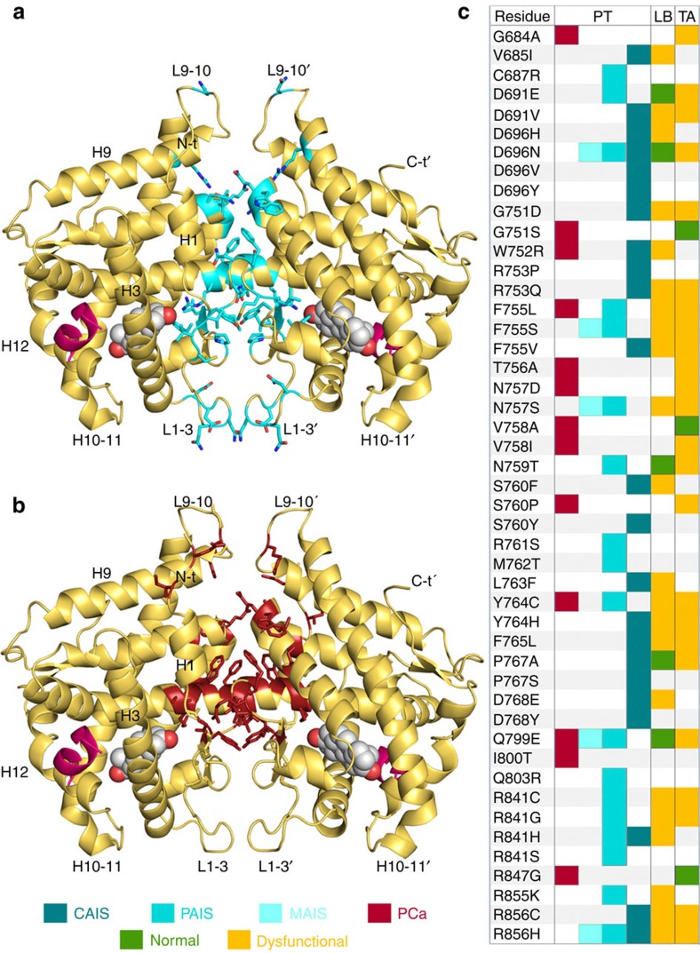
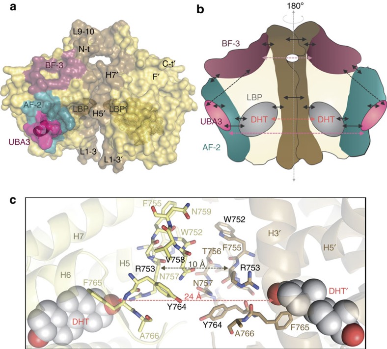
The androgen receptor (AR) plays a crucial role in normal physiology, development and metabolism as well as in the aetiology and treatment of diverse pathologies such as androgen insensitivity syndromes (AIS), male infertility and prostate cancer (PCa). Here we show that dimerization of AR ligand-binding domain (LBD) is induced by receptor agonists but not by antagonists. The 2.15-Å crystal structure of homodimeric, agonist- and coactivator peptide-bound AR-LBD unveils a 1,000-Å2 large dimerization surface, which harbours over 40 previously unexplained AIS- and PCa-associated point mutations. An AIS mutation in the self-association interface (P767A) disrupts dimer formation in vivo, and has a detrimental effect on the transactivating properties of full-length AR, despite retained hormone-binding capacity. The conservation of essential residues suggests that the unveiled dimerization mechanism might be shared by other nuclear receptors. Our work defines AR-LBD homodimerization as an essential step in the proper functioning of this important transcription factor.


Conflict of interest statement

The authors declare no competing financial interests.

Figures

Figure 1
Figure 1. Crystal structure of AR-LBD in complex with UBA3 peptide.
(a) An UBA3 peptide comprising the canonical LxxLL motif binds tightly to AR-LBD. The results of SPR studies conducted in triplicate are shown. (b) Closeup around the AF-2 binding groove with the bound UBA3 peptide shown as a cartoon (pink, with leucine side chains represented as sticks). AR is also depicted as a cartoon with AF-2 and BF-3 binding areas highlighted in brighter blue and magenta, respectively, and the bound DHT moiety in sphere representation. (c,d) Details of the final electron density map. Most relevant AR-LBD residues are represented as sticks and H-bonds with black dotted lines. (c) Closeup showing major interactions across the interface of the core dimer composed by the arbitrarily labelled molecules B (in yellow) and C (in brown). Electron density is shown as either a brown or yellow mesh contoured at 1σ. (d) Closeup showing docking of H6 from peripheral AR-LBD molecule A (pale blue) into the BF-3 pocket of AR-LBD molecule B (yellow). Residues from the peripheral monomer are marked with an asterisk. (e,f) Two views of the AR-LBD crystal structure with the four independent AR-LBD molecules (ad) found in the ASU. Notice that AR-LBD monomers B (yellow) and C (brown) form a symmetrical core dimer, while the two peripheral AR-LBD labeled as (a) shown (teal) and (d) (pale blue) are associated to the BF-3 grooves of (b,c) respectively.
Figure 2
Figure 2. Details of the AR-LBD dimer interface.
(a) Overall structure of the AR-LBD core dimer. The two monomers are depicted as cartoons, with monomer B (yellow) in standard orientation and monomer C in brown; helices and loops are marked. The hormone (dihydrotestosterone, DHT) and the UBA3 peptide are shown as spheres and as a cartoon, respectively. (b) Surface representation of the AR-LBD homodimer shown in the same orientation and coloured yellow and brown as in a. The side chains of residues involved in direct inter-monomer contacts are represented as sticks, coloured according to the monomer they belong to. The DHT moieties are depicted as color-coded spheres (oxygen, red; carbon, yellow or brown). The ‘right' AR-LBD monomer is titled by ∼20° perpendicular to the pseudo twofold axis relating the partners, which results in a slightly asymmetric dimer. (ce) Closeups of the AR-LBD dimer interface highlighting major inter-domain contacts. Residues are shown as color-coded sticks (oxygen, red; nitrogen, blue; carbon, yellow or brown) and labelled. Hydrogen bonding interactions are indicated with black dots. (f) Closeup of the H6 helix from monomer A docking onto the BF-3 pocket of monomer B. Relevant residues are depicted as sticks and H-bonds as black dotted lines. The Tyr774* residues of the peripheral monomers occupy topologically equivalent positions as the outer ring of TRIAC (g) or the benzoic ring of FLF (h). Residues from the peripheral monomers are marked with an asterisk. See also Supplementary Fig. 1.
Figure 3
Figure 3. AR dimerizes in solution through the H5-H5' interface.
SPR analysis of AR-LBD self-association by kinetics (a) or affinity (b). The results of experiments conducted in duplicate are shown along with the respective calculated affinity constants. (c) Closeup of the core dimer interface highlighting the close proximity between the C687 Sγ atoms from both monomers. (d) BMOE-induced cross-linking of AR-LBD. The molecular masses (in kDa) of standard proteins are shown at the left side of the gel (MW). Notice detection of an AR-LBD dimer along with bands corresponding to higher-order aggregates in the presence but not in the absence of the crosslinker. (e,f) Representative MS/MS spectra identifying BMOE-crosslinked peptides that include residues C687 from both monomers. See also Supplementary Fig. 2 and Supplementary Table 1.
Figure 4
Figure 4. Functional characterization of homotypic AR-LBD interactions by FRET.
(a) Schematic representation of the generated fusion proteins. (b) Acceptor photobleaching FRET of N-terminal and C-terminal fusions of AR-LBD shows agonist-induced interactions (DHT, (n=65), T (n=32), and R1881 (n=48), while no interactions were observed without hormone (n=44) or when antagonists (Bic (n=38), Enza (n=46), and OHF (n=44)) were bound to the LBD (mean values and standard error of the mean of at indicated number of cells are shown). Representative confocal images of cells expressing the fusions of AR with EYFP/ECFP in the presence of these compounds are displayed below the bars. (c) Acceptor photobleaching FRET of indicated proteins shows loss of interaction for the AR P767A mutant (n=67) when compared with the WT (n=59), but not for the Y764C mutant (n=63; mean values and s.e.m. of indicated number of cells are shown). Representative confocal images of Hep3B cells transiently expressing the indicated protein in the presence of DHT are displayed below the bars. (d) Binding affinity of the EYFP-AR-LBD fusion protein for the AR agonist mibolerone. (e) Maximal binding of WT and mutant AR for mibolerone (mean values and standard error of the mean of three experiments with three technical replicates each are shown).
Figure 5
Figure 5. Functional validation of the AR-LBD dimer interface.
To investigate the impact of mutations predicted to influence dimerization of the full-length receptor, luciferase reporter and whole-cell competition assays were performed in PC-3 and COS-7 cells, respectively. The mean and s.e.m. of four independent experiments with three technical replicates each are shown for both assays. (a) Transactivation assays were performed with increasing concentrations of DHT (from 0.1 to 10 nM). (b,c) Determination of relative binding affinities for DHT and of maximum binding of mibolerone. (d,e) Western blot analysis of wild-type and mutant AR variants from the experiments shown in panels (ac), respectively.
Figure 6
Figure 6. Mutations associated with AIS and PCa cluster in the AR-LBD dimer interface.
Cartoon representation of the AR-LBD dimer (yellow) with the side chains of all mutated interface residues shown with all their non-hydrogen atoms as sticks, coloured blue for AIS (a) or red for PCa (b). (c) A complete list of AR missense mutations that affect interface residues reported to date, along with their associated phenotypes. Mutations have been taken from the Androgen Receptor Gene Mutations Database (http://androgendb.mcgill.ca/). For a detailed bioinformatics analysis of the predicted impact of these exchanges see Supplementary Fig. 3, Supplementary Notes and Supplementary Tables 3 and 5.
Figure 7
Figure 7. Proposed allosteric communication pathways across the AR-LBD dimer interface.
(a) Surface representation of the AR homodimer. The dimer interface (brown), the AF-2 groove (blue) and the BF-3 pocket (raspberry) are highlighted. Residues that form or line the LBP are shown with a Connolly dot surface and the UBA3 peptide as a pink surface. (b) Schematic representation of the proposed intra- and inter-domain allosteric pathways in AR-LBD. Solid arrows indicate short-range communication networks, while dashed arrows point to long-range interactions. (c) Close-up of the dimer interface highlighting allosteric communication between the LBPs across the dimer interface. The distances between the two R753 residues and the two DHT moieties are given.

Comment in

References

    1. Huang P., Chandra V. & Rastinejad F. Structural overview of the nuclear receptor superfamily: insights into physiology and therapeutics. Annu. Rev. Physiol. 72, 247–272 (2010). - PMC - PubMed
    1. Evans R. M. & Mangelsdorf D. J. Nuclear receptors, RXR & the big bang. Cell 157, 255–266 (2014). - PMC - PubMed
    1. Vanderschueren D. et al.. Sex steroid actions in male bone. Endocr. Rev. 35, 906–960 (2014). - PMC - PubMed
    1. Gottlieb B., Beitel L. K., Nadarajah A., Paliouras M. & Trifiro M. The androgen receptor gene mutations database: 2012 update. Hum. Mutat. 33, 887–894 (2012). - PubMed
    1. Hughes I. A. et al.. Androgen insensitivity syndrome. Lancet 380, 1419–1428 (2012). - PubMed

Publication types

MeSH terms