. 2017 Mar;35(3):264-272.
doi: 10.1038/nbt.3801. Epub 2017 Feb 6.

Gene therapy restores auditory and vestibular function in a mouse model of Usher syndrome type 1c

Affiliations

Gene therapy restores auditory and vestibular function in a mouse model of Usher syndrome type 1c

Bifeng Pan et al. Nat Biotechnol. 2017 Mar.

Abstract

Because there are currently no biological treatments for hearing loss, we sought to advance gene therapy approaches to treat genetic deafness. We focused on Usher syndrome, a devastating genetic disorder that causes blindness, balance disorders and profound deafness, and studied a knock-in mouse model, Ush1c c.216G>A, for Usher syndrome type IC (USH1C). As restoration of complex auditory and balance function is likely to require gene delivery systems that target auditory and vestibular sensory cells with high efficiency, we delivered wild-type Ush1c into the inner ear of Ush1c c.216G>A mice using a synthetic adeno-associated viral vector, Anc80L65, shown to transduce 80-90% of sensory hair cells. We demonstrate recovery of gene and protein expression, restoration of sensory cell function, rescue of complex auditory function and recovery of hearing and balance behavior to near wild-type levels. The data represent unprecedented recovery of inner ear function and suggest that biological therapies to treat deafness may be suitable for translation to humans with genetic inner ear disorders.


Conflict of interest statement

Competing Financial Interests Statement

A patent #00633-0203P01 on “Materials and methods for delivering nucleic acids to cochlear and vestibular cells” has been deposited by L.H.V., G.S.G. & J.R.H. The Anc80L65 vector is patented by L.H.V., patent #WO/2015/054653 – “Methods of predicting ancestral virus sequence and uses thereof. L.H.V. is co-founder and SAB member of GenSight Biologics and consultant to various gene therapy companies. LHV receives research support from Selecta Biosciences and Lonza Houston on Anc-AAV development and discovery.

Figures

Figure 1
Figure 1. Scanning electron microscopy of the organ of Corti in Ush1c c.216G>A mice at P8
(a–f) Basal, middle and apical regions of the organ of Corti were imaged in P8 c.216GA (n = 3 mice) and c.216AA (n = 4 mice) mice. OHC and IHC hair bundles were preserved in heterozygous mice but some hair bundles appeared disorganized along the organ of Corti in homozygous 216AA mice. (g–l) High magnification images revealed fragmented and disorganized bundles with disruptions in the staircase array in many but not all OHC (g–h) and IHCs (i–j). Examples of OHC hair bundles imaged in the middle region of the organ at P8 illustrate a preserved (k) and a disorganized hair bundle (l) present in the same preparation. Stars indicate preserved hair bundles; arrowhead, disorganized hair bundles; and arrows, wavy IHC bundles. Scale bars low mag.: 5 μm (a–f); high mag.: 2 μm (g), 3 μm (h), 2 μm (i–j) and 1 μm (k,l).
Figure 2
Figure 2. Mechanotransduction in hair cells of Ush1c c.216G>A neonatal mice
(a–d) The permeable styryl dye FM1-43 was used to assess the presence of open transduction channels in hair cells of c.216GA and c.216AA mice. In the organ of Corti, FM uptake was reduced in sensory hair cells of c.216AA mice at P4 (a–b, mid base). Note that IHC FM1-43 fluorescence appears dimmer as IHCs are in a different focal plane. Left: DIC, Right: FM1-43; Scale bar 10μm. In the utricle, FM1-43 uptake was restricted to the extra-striola region in c.216AA mutants at P6 (c; scale bar 50μm) while utricular hair cells retained gross normal bundle morphology as assessed by DIC (d; scale bar 10μm). The white lines on panel c and d delineate the striola (no uptake) and extra-striola regions (uptake). Experiment was repeated three times. (e–h) Mechanotransduction was assessed in OHCs, IHCs and VHCs in neonatal c.216GA and c.216AA mice (number of mice recorded from respectively are: n= 7, 6 for OHCs, n= 2, 4 for IHCs and n= 2, 6 for VHCs, number of cells are indicated above the bar graph; Holding potential: −60mV). Representative transduction currents (e), their associated current/displacement plots fitted with a second order Boltzmann function (f) and average peak transduction current are plotted (g–h). In the cochlea, recordings were obtained in the middle and mid-apical turn of the organ at P3-P6. In the utricle transduction currents were recorded from VHCs of the extra-striola (E.S.) and striola (S.) region between P5 and P7 (e–f). While hair bundles appeared well preserved under DIC, smaller average transduction currents were evoked in c.216AA mutants (h). Average peak transduction was significantly different between the two genotypes in OHCs, IHCs and VHCs (***P < 0.01, One-way ANOVA).
Figure 3
Figure 3. Expression and localization of fluorescently labeled harmonin in tissues exposed to adeno-associated viral vectors in vitro and in vivo
(a–c) Acutely dissected P0-P1 inner ear tissue were exposed to AAV2/1 vectors for 24h, kept in culture for 7 to 8 days before being fixed, counterstained (Alexa Fluor phalloidin, Invitrogen) and imaged with a Zeiss LSM confocal microscope. A large number of sensory hair cells were infected in wild-type utricle and expression of harmonin-b1 fused to EGFP was evident in most hair cells with specific localization at the apex of the sensory hair bundle (a, scale bar: 10 μm- upper panels; 5μm- lower panels). Similarly expression of EGFP::harmonin-b1 was evident at the tip of the stereocilia in OHCs and IHCs of c.216AA and wild-type mice (b, scale bar: 10 μm; c, scale bar: 3μm). When 1 μl AAV2/1.CMV.EGFP::harmonin-b1 vectors were injected at P1, EGFP signal was detected in some IHCs and OHCs at P60 in the left injected ear (d, scale bar: 30 μm). Exogenous tdTomato::harmonin-a1 was detected in the cell body of IHCs and OHCs in P7 organotypic cultures exposed to AAV2/1.CMV.tdTomato::harmonin-a1 for 24 h at P0 (e, scale bar: 5μm). Some harmonin-a1 puncta were colocalized with CTBP2 (blue; mouse anti-CTBP2 1/200, BD bioscience) in particular at the base of the sensory cells presumably near the ribbon synapse (arrowheads). No expression was observed in the stereociliary bundle.
Figure 4
Figure 4. Recovery of mechanotransduction in hair cells of mice injected with Anc80L65 harmonin vectors
(a–c) Mechanotransduction currents were recorded in IHCs of c.216AA uninjected control mice (n=8 cells, one mouse) and c.216AA mice injected at P1 with AAV2/Anc80L65.CMV.harmonin-b1 (0.8 μl, 1.9×10^12 gc/ml, n=15 cells, one mouse) or a combined injection of the AAV2/Anc80L65.CMV.harmonin-a1 (1.7×10^12 gc/ml) and AAV2/Anc80L65.CMV.harmonin-b1 (0.5 μl + 0.5 μl, n=7 cells, one mouse). Tissue was extracted at P5-P6, before the cochlea became ossified, and was maintained in culture for 10 days. (9 to 10 DIV). While small mechanotransduction currents could be induced by hair bundle stimulations of c.216AA mice, larger currents were evoked in c.216AA mice injected with vectors driving harmonin-b1 or dual harmonin-a1 and -b1 expression (a). Corresponding I/X curve for each dataset and double Boltzmann fitting function. Respective maximal mechanotransduction current Imax= 102.1 pA (c.216AA); 424.3 pA (c.216AA + harmonin-b1) and 341.1 pA (c.216AA + harmonin-a1&-b1) (b). Average responses (Mean ± S.D.) show significant recovery of transduction (***P < 0.001) for harmonin-b1 and harmonin-a1 +-b1 injected relative to uninjected mice. Average transduction currents were not significantly different in harmonin-b1 injected mice and c.216GA control mice (N.S. P>0.5). Recovery of mechanotransduction was also not significantly improved when harmonin-a and harmonin-b were combined. (c), one-way ANOVA.
Figure 5
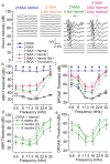
Figure 5. ABR and DPOAE threshold recovery in mice injected at P1 with AAV2/Anc80L65.CMV.harmonin-b1
(a) Representative ABR responses for 16 kHz tones in 6 weeks old c.216AA control mice and c.216AA mice injected at P1 via RWM injection of vectors encoding harmonin-a1(0.8 μl, 1.7×10^12 gc/ml), harmonin-b1 (0.8 μl, 1.9×10^12 gc/ml) or a combination of the two (0.5 μl + 0.5 μl). Recovered ABR thresholds near 30 dB were measured in mice injected with harmonin-b1 alone or harmonin-a1 and b1 together. (b) Mean ABR responses obtained for: c.216 AA (n=13); c.216GA (n=12); c.216GA + trunc-harmonin (n=4); c.216AA + harmonin-a1 (n=12); c.216AA + harmonin-b1 (n=19 with rescued ABR thresholds < 80 dB of 25 tested); c.216AA + harmonin-a1&-b1 (n=6 rescued with ABR thresholds <80 dB/11 tested). Mean ± S.E, continuous lines. Dotted lines: ABR thresholds for the entire frequency range in mice whose 16 kHz recordings are shown in panel a. (c) Mean DPOAEs responses obtained for: c.216AA (n=13); c.216GA (n=12); c.216GA + trunc-harmonin (n=4); c.216AA + harmonin-a1 (n=12); c.216AA + harmonin-b1 (n=15 rescued with DPOAE thresholds <70 dB/25 tested); c.216AA + harmonin-a1&-b1 (n=4 rescued with DPOAEs <70 dB/11 tested). Mean ± S.E, continuous lines. Dotted lines: DPOAEs thresholds for the four mice whose recordings are illustrated in panel A. Arrows indicate that the thresholds are higher than the maximal stimulus level tested. (d–e) ABRs and DPOAEs responses obtained at 6 weeks and 3 months in eight mice that showed initial ABR thresholds under or equal to 45 dB SPL. Seven of the eight mice were kept for 6 months (one taken for histology) and had ABRs and DPOAEs assessed (dotted line). Mean ± S.E. While ABRs and DPOAEs thresholds shifts were evident over the first three month, hearing rescue was still prominent at 6 months of age in the lower frequency range.
Figure 6
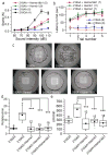
Figure 6. Startle response, rotarod performance and open field behavior recovery in mice injected at P1 with AAV2/Anc80L65.CMV.harmonin-a1 and AAV2/Anc80L65.CMV.harmonin-b1
(a) Startle response to white noise stimuli was recorded in 6 weeks old control c.216GA, c.216AA mice and injected mice. Partial but significant startle rescue was evident in mice injected with harmonin-b1 but not harmonin-a1 (data overlap with control c.216AA mice). Averages ± S.E. are shown for all mice tested. Statistical analysis: 216AA relative to 216AA + Harm a1&b1: p=0.008 at 100db; p= 0.005 at 110dB; 216AA relative to 216AA + Harm b1: p= 0.007 at 100dB; 0.001 at 110dB, Student t-test. (b) Rotarod performance was recorded between 4 and 6 weeks in control c.216GA, c.216AA and injected mice. Full recovery was observed in all mice tested that were injected with harmonin-b1 and harmonin-a1/b1. No recovery was observed for harmonin-a1 alone. Averages ± S.E. are shown for all mice tested. (c–e) Open field observations were performed in 42 cm wide arena for 5 min in 6 weeks old control c.216GA, c.216AA and injected mice. Representative tracks over 2.5 min are shown (c). While c.216AA mice explore the entire field, and perform repetitive full-body rotations, c.216AA mice injected at P1 with harmonin-a1, harmonin-b1 or the combination of the two vectors demonstrate normal behavior similar to their heterozygous c.216GA. Mean ± S.D. for the number of rotations (d) and distance covered (e) per minute for all mice tested. Significant recovery ***P < 0.001 was observed between the uninjected and injected mice. Statistical analysis by one-way ANOVA.
Figure 7
Figure 7. Scanning electron microscopy of the organ of Corti in mice injected with AAV2/Anc80L65.CMV.harmonin-b1
Basal, Middle and Apical regions of the organ of Corti were imaged at six weeks in c.216GA (a–d), c.216AA (e–h) and c.216AA (i–l, n–p) mice injected at P1 (RMW injection 0.8 μl AAV2/Anc80L65.CMV.harmonin-b1). OHC and IHC hair bundles were preserved in c.216GA mice but appeared disorganized along the organ of Corti in c.216AA mice. Noticeable hair cell loss (asterisk) and hair bundle disorganization was observed in c.216AA mice with more pronounced degeneration in the basal end of the organ. Hair bundles of c.216AA mice lacked normal stereocilia rows. The shorter rows appeared to be retracted while the tallest rows were maintained in c.216AA mice (arrow). While hair cell loss and bundle disorganization were still evident in rescued c.216AA mice, hair cell survival was noticeably higher in the basal and middle regions of the Organ. Hair cell counts are summarized in the bar graph. A total of 1824 cells were counted in c.216AA mice (4 ears) and 792 in rescued c.216AA mice (2 ears). Mean ± S.E. High magnification imaging reveals rescue of the staircase array in injected c.216AA mice (arrow) in many but not all cells (arrowhead). Scale bar low magnification: 5 μm; high magnification: 1 μm.

Comment in

  • Hearing in the mouse of Usher.
    Brigande JV. Brigande JV. Nat Biotechnol. 2017 Mar 7;35(3):216-218. doi: 10.1038/nbt.3815. Nat Biotechnol. 2017. PMID: 28267741 Free PMC article.

References

    1. Chien WW, et al. Gene Therapy Restores Hair Cell Stereocilia Morphology in Inner Ears of Deaf Whirler Mice. Mol Ther. 2016;24:17–25. - PMC - PubMed
    1. Askew C, et al. Tmc gene therapy restores auditory function in deaf mice. Sci Transl Med. 2015;7:295ra108. - PMC - PubMed
    1. Akil O, et al. Restoration of hearing in the VGLUT3 knockout mouse using virally mediated gene therapy. Neuron. 2012;75:283–293. - PMC - PubMed
    1. Boughman JA, Vernon M, Shaver KA. Usher syndrome: definition and estimate of prevalence from two high-risk populations. J Chronic Dis. 1983;36:595–603. - PubMed
    1. Smith RJ, et al. Clinical diagnosis of the Usher syndromes. Usher Syndrome Consortium. Am J Med Genet. 1994;50:32–38. - PubMed

MeSH terms