. 2017 Feb 6:7:41916.
doi: 10.1038/srep41916.

Restenosis Inhibition and Re-differentiation of TGFβ/Smad3-activated Smooth Muscle Cells by Resveratrol

Affiliations

Restenosis Inhibition and Re-differentiation of TGFβ/Smad3-activated Smooth Muscle Cells by Resveratrol

Yichen Zhu et al. Sci Rep. .

Abstract

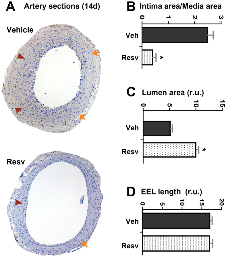
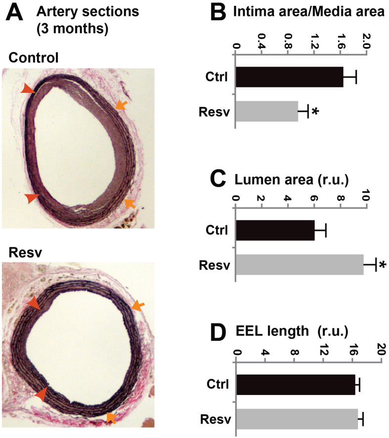
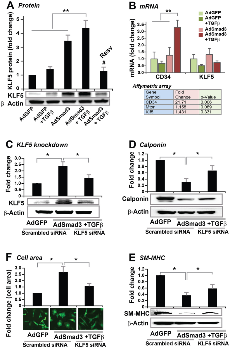
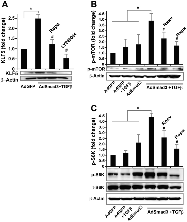
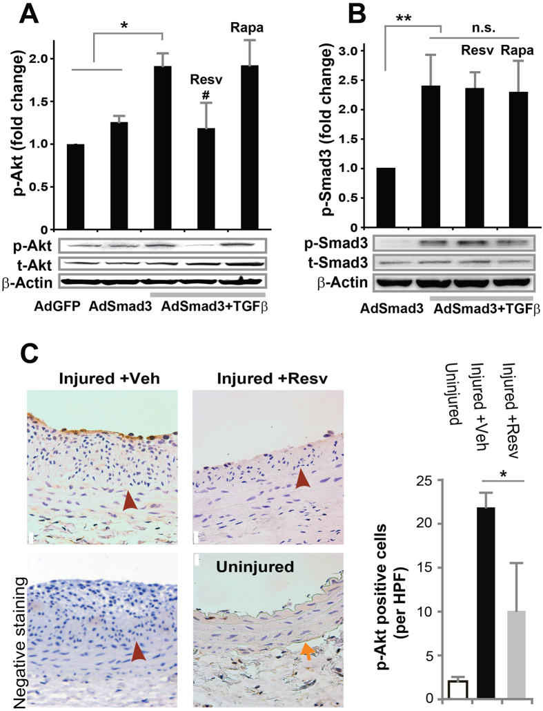
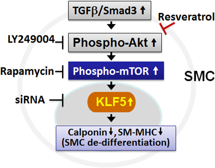
To date, there is no periadventitial drug delivery method available in the clinic to prevent restenotic failure of open vascular reconstructions. Resveratrol is a promising anti-restenotic natural drug but subject to low bioavailability when systemically administered. In order to reconcile these two prominent issues, we tested effects of periadventitial delivery of resveratrol on all three major pro-restenotic pathologies including intimal hyperplasia (IH), endothelium impairment, and vessel shrinkage. In a rat carotid injury model, periadventitial delivery of resveratrol either via Pluronic gel (2-week), or polymer sheath (3-month), effectively reduced IH without causing endothelium impairment and vessel shrinkage. In an in vitro model, primary smooth muscle cells (SMCs) were stimulated with elevated transforming growth factor (TGFβ) and its signaling protein Smad3, known contributors to IH. TGFβ/Smad3 up-regulated Kruppel-like factor (KLF5) protein, and SMC de-differentiation which was reversed by KLF5 siRNA. Furthermore, TGFβ/Smad3-stimulated KLF5 production and SMC de-differentiation were blocked by resveratrol via its inhibition of the Akt-mTOR pathway. Concordantly, resveratrol attenuated Akt phosphorylation in injured arteries. Taken together, periadventitial delivery of resveratrol produces durable inhibition of all three pro-restenotic pathologies - a rare feat among existing anti-restenotic methods. Our study suggests a potential anti-restenotic modality of resveratrol application suitable for open surgery.


Conflict of interest statement

The authors declare no competing financial interests.

Figures

Figure 1
Figure 1. Effect of two-week resveratrol treatment on neointima formation, vessel size, and lumen size of rat carotid arteries following balloon injury.
(A) Representative stained sections from arteries treated with vehicle (DMSO) and resveratrol respectively and collected on day 14 after injury. Arrows indicate external elastic lamina (EEL); arrowheads mark internal elastic lamina (IEL). (BD) Quantified data of IH (ratio of intima/media area), lumen area, and arterial remodeling (EEL length), respectively. Each bar represents a mean (±SEM) of sections from 5 animals, *P < 0.05 compared to vehicle control.
Figure 2
Figure 2. Effect of resveratrol treatment on re-endothelialization and cell apoptosis of rat carotid arteries after balloon injury.
Rat carotid arteries were treated with either vehicle or resveratrol and collected on day 14 after injury. Arrows indicate external elastic lamina (EEL); arrowheads mark internal elastic lamina (IEL). (A) Immunostaining of CD31. Quantification: mean ± SEM; n = 3–5 animals; *P < 0.05 compared to vehicle control. (B) Immunostaining of cleaved caspase-3. Scale bar = 20 μm. A placenta sample serves as positive control.
Figure 3
Figure 3. Effect of three-month resveratrol treatment on neointima formation, vessel size, and lumen size of rat carotid arteries following balloon injury.
(A) Representative van Gieson-stained sections from arteries treated respectively with control PCL sheaths (no resveratrol) and resveratrol-loaded sheaths and collected at 3 months after injury. Arrows indicate external elastic lamina (EEL); arrowheads mark internal elastic lamina (IEL). (BD) Quantified data of IH (ratio of intima/media area), lumen area, and arterial remodeling (EEL length), respectively. Each bar represents a mean (±SEM) of sections from 4–5 animals, *P < 0.05 compared to vehicle control.
Figure 4
Figure 4. Inhibitory effect of resveratrol on TGFβ/Smad3-stimulated SMC de-differentiation in vitro.
Rat aortic SMCs were infected with AdGFP (control) or AdSmad3 followed by pre-incubation with vehicle (DMSO) or resveratrol (50 μM) or rapamycin (200 nM) for 2 h, and then treated with solvent (for AdGFP control) or TGFβ1 (5 ng/mL) for 24 h. Cells were then used for Western blotting of calponin (A) or SM-MHC (B), or staining with Calcein for cell area measurement (C). Quantification: mean ± SEM of three independent experiments; normalization to β-actin; *P < 0.05 compared to AdGFP control; #P < 0.05 compared to AdSmad3 + TGFβ1 without pre-incubation with a drug.
Figure 5
Figure 5. Inhibitory effect of resveratrol on TGFβ/Smad3-stimulated KLF5 protein production in vitro.
Rat aortic SMCs were infected with AdGFP (control) or AdSmad3 followed by pre-incubation with vehicle (DMSO) or resveratrol (50 μM) for 2 h, and then treated with solvent or TGFβ1 (5 ng/mL) for 24 h. Cells were then collected for Western blotting analysis (A) or RT-PCR (B). Microarray data were retrieved from our recent report. Quantification: mean ± SEM of three independent experiments; normalization to β-actin; **P < 0.05 compared to any of the first 3 conditions; #P < 0.05 compared to AdSmad3 + TGFβ1 without pre-incubation with resveratrol. To determine the effect of KLF5 knockdown on TGFβ/Smad3-stimulated SMC de-differentiation, rat aortic SMCs were infected with AdGFP (control) or AdSmad3 and then transfected with scrambled or KLF5-specific siRNA for 6 h followed by treatment with solvent (control) or TGFβ1 (5 ng/mL) for 24 h. Cells were then used for Western blotting of KLF5 (C), calponin (D) or SM-MHC (E), or staining with Calcein for cell area measurement (F). Quantification: mean ± SEM of three independent experiments; normalization to β-actin; *P < 0.05 compared to AdSmad3 + TGFβ1 with scrambled siRNA (the middle bar).
Figure 6
Figure 6. Inhibitory effect of resveratrol on TGFβ/Smad3-stimulated mTOR activation in vitro.
Rat aortic SMCs were infected with AdGFP (control) or AdSmad3 followed by pre-incubation with vehicle (DMSO), resveratrol (50 μM), rapamycin (200 nM), or LY249004 (20 μM) for 2 h, and then treated with solvent or TGFβ1 (5 ng/mL) for 24 h. Cells were then collected for Western blotting analysis of KLF5 (A), p-mTOR (B) or p-S6K (C). Quantification: mean ± SEM of three independent experiments; normalization to β-actin or to total protein; *P < 0.05 compared to any of the first 3 conditions; #P < 0.05 compared to AdSmad3 + TGFβ1 without pre-incubation with a drug.
Figure 7
Figure 7. Inhibitory effect of resveratrol on TGFβ/Smad3-stimulated Akt activation in vitro and in vivo in balloon-injured rat carotid arteries.
(A,B) Rat aortic SMCs were infected with AdGFP (control) or AdSmad3 followed by pre-incubation with vehicle (DMSO), resveratrol (50 μM), or rapamycin (200 nM) for 2 h, and then treated with solvent or TGFβ1 (5 ng/mL). For Western blotting analyses of p-Akt (A) and p-Smad3 (B), cells were collected after TGFβ1 treatment for 2 h and 0.5 h, respectively. Quantification: mean ± SEM of three independent experiments; normalization to total Akt (t-Akt) or to total Smad3 (t-Smad3); *P < 0.05 compared to each of the first 2 conditions; **P < 0.05 compared to AdSmad3 only; #P < 0.05 compared to AdSmad3 + TGFβ1 without pre-incubation with a drug; n.s., not significant. (C). Rat carotid arteries were treated with either vehicle or resveratrol and collected on day 14 after angioplasty. Shown on the left are representative sections of p-Akt Immunostaining. Arrows indicate external elastic lamina (EEL); arrowheads mark internal elastic lamina (IEL). Scale bar = 20 μm. Negative staining: IgG instead of a primary antibody was used. Quantification: mean ± SEM of 4–5 animals; *P < 0.05 compared to vehicle control; HPF, high power field.
Figure 8
Figure 8. A schematic of resveratrol-targeted pathway in rat vascular smooth muscle cells.

Similar articles

Cited by

References

    1. Goel S. A., Guo L. W., Liu B. & Kent K. C. Mechanisms of post-intervention arterial remodelling. Cardiovascular research 96, 363–371 (2012). - PMC - PubMed
    1. Ichimoto E. et al.. Mechanism of edge restenosis after sirolimus-eluting stent implantation. J Invasive Cardiol 24, 55–57 (2012). - PubMed
    1. Inoue T. et al.. Vascular inflammation and repair: implications for re-endothelialization, restenosis, and stent thrombosis. JACC. Cardiovascular interventions 4, 1057–1066 (2011). - PMC - PubMed
    1. Holy E. W. et al.. PI3K/p110alpha inhibition selectively interferes with arterial thrombosis and neointima formation, but not re-endothelialization: potential implications for drug-eluting stent design. European heart journal 35, 808–820 (2014). - PubMed
    1. Guo L. W. et al.. Halofuginone stimulates adaptive remodeling and preserves re-endothelialization in balloon-injured rat carotid arteries. Circ Cardiovasc Interv 7, 594–601 (2014). - PMC - PubMed

Publication types

MeSH terms

LinkOut - more resources