. 2017 Feb 6:7:41823.
doi: 10.1038/srep41823.

Wnt7a Inhibits IL-1β Induced Catabolic Gene Expression and Prevents Articular Cartilage Damage in Experimental Osteoarthritis

Affiliations

Wnt7a Inhibits IL-1β Induced Catabolic Gene Expression and Prevents Articular Cartilage Damage in Experimental Osteoarthritis

Averi L Gibson et al. Sci Rep. .

Abstract

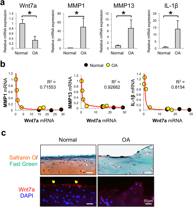
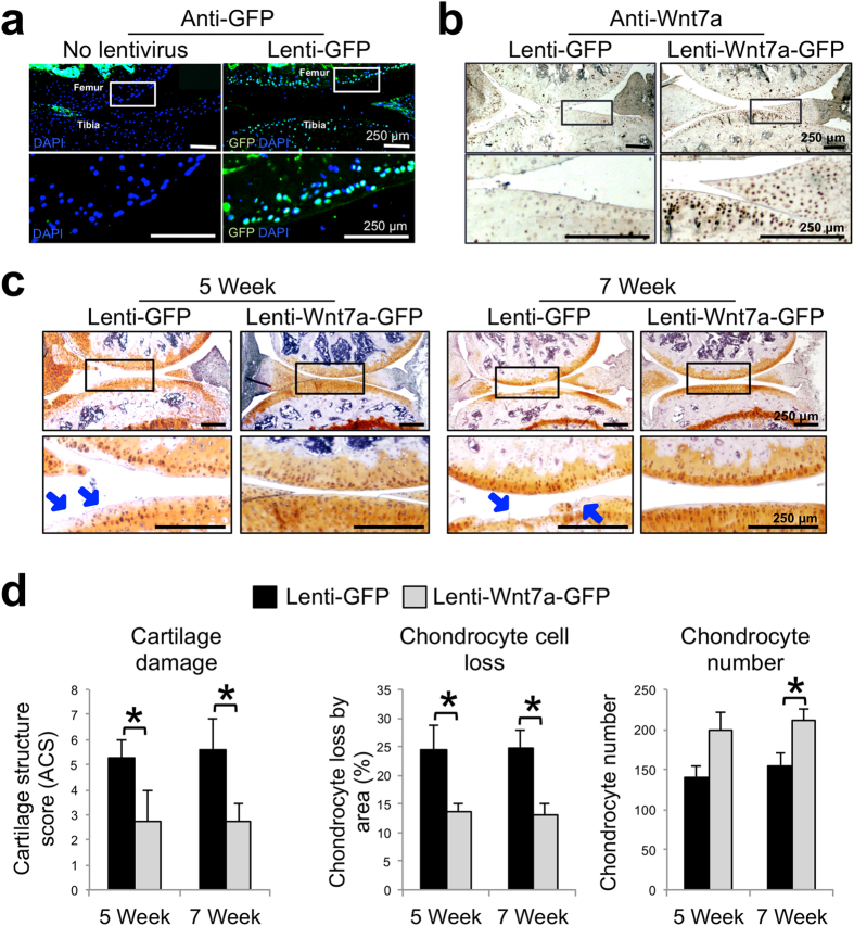
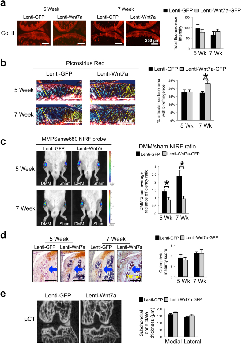
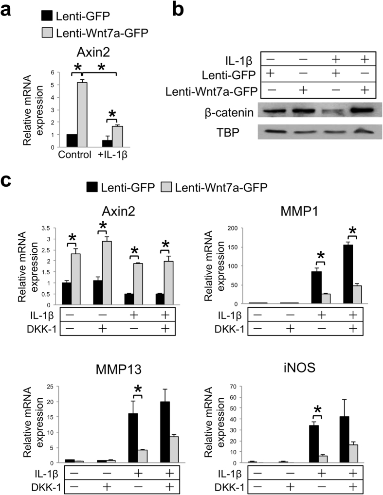
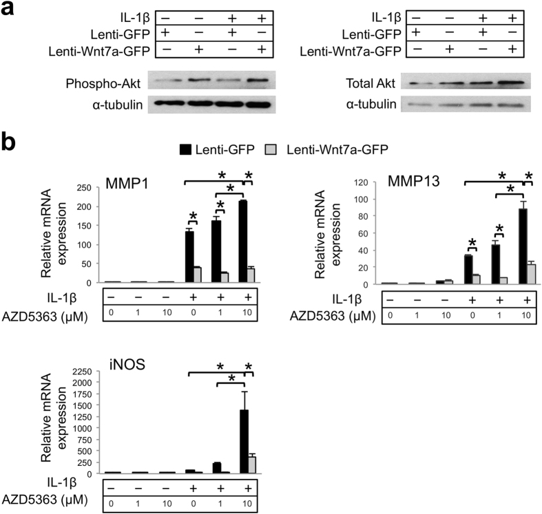
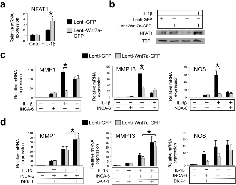
Wnt7a is a protein that plays a critical role in skeletal development. However, its effect on cartilage homeostasis under pathological conditions is not known. In this study, we found a unique inverse correlation between Wnt7a gene expression and that of MMP and IL-1β in individual human OA cartilage specimens. Upon ectopic expression in primary human articular chondrocytes, Wnt7a inhibited IL-1β-induced MMP and iNOS gene expression. Western blot analysis indicated that Wnt7a induced both canonical Wnt signaling and NFAT and Akt non-canonical signaling. Interestingly, inhibiting the canonical and Akt pathway did not affect Wnt7a activity. However, inhibiting the NFAT pathway impaired Wnt7a's ability to inhibit MMP expression, suggesting that Wnt7a requires NFAT signaling to exert this function. In vivo, intraarticular injection of lentiviral Wnt7a strongly attenuated articular cartilage damage induced by destabilization of the medial meniscus (DMM) OA-inducing surgery in mice. Consistently, Wnt7a also inhibited the progressive increase of joint MMP activity in DMM animals. These results indicate that Wnt7a signaling inhibits inflammatory stimuli-induced catabolic gene expression in human articular chondrocytes and is sufficient to attenuate MMP activities and promote joint cartilage integrity in mouse experimental OA, demonstrating a novel effect of Wnt7a on regulating OA pathogenesis.


Conflict of interest statement

The authors declare no competing financial interests.

Figures

Figure 1
Figure 1. Wnt7a mRNA expression is reduced in human OA and inversely correlated with catabolic gene expression.
(a) RT-PCR analysis of Wnt7a and MMP1, MMP3 and IL-1β gene expression in tibial cartilage specimens isolated from normal cadaveric and OA patients. The age and sex of human cartilage specimens used were: normal (n = 4): 84/M, 75/M, 65/F, and 49/F. OA specimens (n = 6): 53/F, 63/F, 65/F, 72/M, 73/F, 73/M. (b) Correlation analysis of Wnt7a mRNA expression with catabolic gene expression in normal cadaveric and OA patient samples. Data points from the six OA samples scatter toward the left of the graphs (yellow dots), and those from the four normal samples are scattered to the right of the graphs (black dots), with two of the normal sample points overlapping with one another. (c) Normal and OA human cartilage samples stained with safranin O/fast green and an anti-Wnt7a antibody. DAPI was used to visualize the nuclei. Diffuse staining was observed in both normal and OA cartilage, but strong Wnt7a protein expression was detected on the articular surface (arrows) in normal cartilage. Experiments were performed in triplicates with technical duplicates. A student’s t-test was used for evaluating the statistical significance of normal vs. OA gene expression. All correlation analyses were performed using a Spearman correlation and were statistically significant (p < 0.05). Data are shown as mean ± SEM. *p < 0.05.
Figure 2
Figure 2. Wnt7a ectopic expression reduces IL-1β induced upregulation of OA-related genes and NF-κB activity in vitro.
(a) Representative images of Wnt7a IF analysis of normal human articular chondrocytes (nHACs) after infection with lenti-GFP or lenti-Wnt7a-GFP. Wnt7a immunofluorescence images were overlaid with GFP expression. Percentage of cells positive for lenti-GFP or lenti-Wnt7a-GFP was quantified based on GFP production, which were 81.4% and 78.8%, respectively. (b) RT-PCR analysis of nHACs gene expression after infection with lenti-GFP or lenti-Wnt7a-GFP with or without IL-1β (5 ng/mL). Lenti-Wnt7a-GFP viral infection significantly reduced IL-1β upregulation of Collagen X, IL-1β, iNOS, and several MMPs. A student’s t-test was used for evaluating the statistical significance between the gene expression of lenti-GFP and lenti-Wnt7a cells under either control or IL-1β conditions. (c) NF-κB luciferase assay of NF-κB activity after infection with lenti-GFP or lenti-Wnt7a-GFP and treatment with or without IL-1β (5 ng/mL). Relative luciferase activity in reference to Renilla luciferase internal control is shown. Experiments were conducted with biological triplicates and repeated at least three times. Analysis of variance (ANOVA) with post-hoc tests was used for evaluating the statistical significance of the GFP vs. Wnt7a and control vs. IL-1β luciferase activity. All data are shown as mean ± SEM. *p < 0.05.
Figure 3
Figure 3. Wnt7a ectopic expression prevents cartilage damage in the DMM OA model in vivo.
(a) Representative images of GFP immunofluorescence (IF) analysis of mouse knee joints 5 weeks post DMM surgery with or without lenti-GFP joint infection. Rectangles denote areas shown in higher magnification. Both the superficial and deeper zones of the articular cartilage were infected. (b) Representative images of Wnt7a IF analysis in mouse knees injected with lenti-GFP or lenti-Wnt7a-GFP at 5 weeks post DMM surgery. Rectangles denote areas shown in higher magnification. Wnt7a IF confirmed ectopic expression of Wnt7a. Quantification analysis based on GFP and Wnt7a expression from at least four independent images for each treatment showed the percentage of tibial articular chondrocytes positive for lenti-GFP or lenti-Wnt7a-GFP was 55.1% and 57.7% respectively. (c) Representative images showing safranin O/hematoxylin stained joint sections at 5 or 7 weeks post DMM surgery in mouse knees injected with lenti-GFP or lenti-Wnt7a-GFP. Rectangles denote areas shown in higher magnification. Arrows denote areas of articular cartilage damage. Lenti-GFP control knees showed significantly more cartilage damage compared to knees ectopically expressing Wnt7a. (d) Cartilage structure scoring (ACS), chondrocyte loss and chondrocyte numbers in the tibia were quantified at 5 or 7 weeks post DMM in mouse knees injected with lenti-GFP or lenti-Wnt7a-GFP. n = 6 mice/group with 9 sections/joint used for blind scoring. Nonparametric statistical analyses were used for evaluating the statistical significance of the histological scoring systems at each time point. Data are shown as mean ± SEM. *p < 0.05.
Figure 4
Figure 4. Wnt7a reduces joint MMP activity during OA development, but has no effect on subchondral bone.
(a) Representative images of Collagen II IHC analysis 5 or 7 weeks post DMM surgery in mouse knees injected with lenti-GFP or lenti-Wnt7a-GFP. Quantification analysis based on total Collagen II fluorescence of the femur and tibia from at least 4 sections/treatment indicated there were no significant differences among groups. (b) Representative images and semi-quantitative analysis of picrosirius red staining showing collagen fiber thickness and organization of articular cartilage from knee joints at 5 or 7 weeks post DMM surgery, as viewed under polarized light. Areas used for subsequent quantification of birefringence signals are delineated by dashed lines. (c) Representative in vivo near infrared fluorescence (NIRF) images taken two hours after 4 μL intraarticular injection of MMPSense680 in each knee. Each lenti-GFP and lenti-Wnt7a-GFP-infected mouse was imaged serially at 5 and 7 weeks post surgery. The average radiance efficiency emitted from each knee was quantified from the collected NIRF images and the DMM average radiance efficiency was divided by the sham average radiance efficiency to internally calibrate each signal. Quantification analysis demonstrated that mice ectopically expressing Wnt7a in the joint have reduced NIRF signals compared to lenti-GFP controls. (d) Representative histological images of osteophyte development with safraninO/hematoxylin staining and scoring of osteophyte maturity at 5 or 7 weeks post DMM surgery in mouse knees injected with lenti-GFP or lenti-Wnt7a-GFP. Arrows indicate osteophytes. Osteophyte maturity was quantified. (e) Representative micro-CT cross-sections of the knee joint and quantification of medial and lateral tibial subchondral bone plate thickness at 7 weeks post DMM surgery in mouse knees injected with lenti-GFP or lenti-Wnt7a-GFP. Micro-CT quantification demonstrated no differences between the lenti-GFP and lenti-Wnt7a-GFP groups. n = 7 mice/group for NIRF imaging and 6 mice/group for microCT imaging and histology. A student’s t-test was used for evaluating the statistical significance between GFP and Wnt7a mice at each individual time point. All data are shown as mean ± SEM. *p < 0.05.
Figure 5
Figure 5. Inhibition of canonical Wnt signaling does not diminish Wnt7a’s inhibition of IL-1β activity in human chondrocytes.
(a) RT-PCR analysis of Axin2 mRNA expression in nHACs after infection with lenti-GFP or lenti-Wnt7a-GFP, and cultured with or without IL-1β (5 ng/mL). (b) Western blot analysis of nuclear β-catenin in nHACs after infection with lenti-GFP or lenti-Wnt7a-GFP in the presence or absence of IL-1β (5 ng/mL). TATA-binding protein (TBP) served as a loading control. Original films for cropped images can be found in the supplementary information file. (c) RT-PCR analysis of Axin2, MMP1, MMP13 and iNOS gene expression after treatment with DKK-1 (250 ng/mL), on nHACs infected with lenti-GFP or lenti-Wnt7a-GFP, and cultured with or without IL-1β (5 ng/mL). Each experiment had three biological repeats/treatment, and at least three independent experiments were performed. A student’s t-test was used for evaluating the statistical significance between the gene expression of lenti-GFP and lenti-Wnt7a cells under each individual experimental condition. All data are shown as mean ± SEM. *p < 0.05.
Figure 6
Figure 6. Inhibition of Akt signaling does not diminish Wnt7a’s inhibition of IL-1β activity in human chondrocytes.
(a) Western blot analysis of Akt protein expression after nHACs were infected with lenti-GFP or lenti-Wnt7a-GFP and cultured with or without IL-1β (5 ng/mL). Results demonstrated that phosphorylated Akt was upregulated with Wnt7a ectopic expression compared to lenti-GFP controls. Original films for cropped images can be found in the supplementary information file. (b) RT-PCR analysis of MMP1, MMP13 and iNOS gene expression after treatment with Akt inhibitor AZD5363 (1 or 10 uM) on nHACs infected with lenti-GFP or lenti-Wnt7a-GFP and cultured in the presence of absence of IL-1β (5 ng/mL). Each experiment had three biological repeats/treatment, and at least three independent experiments were performed. Analysis of variance (ANOVA) with post-hoc tests was used for evaluating the statistical significance between the gene expression of lenti-GFP and lenti-Wnt7a cells across all of experimental conditions. All data are shown as mean ± SEM. *p < 0.05.
Figure 7
Figure 7. Inhibition of NFAT signaling attenuates the effect of Wnt7a on MMP inhibition in human chondrocytes.
(a) RT-PCR analysis of NFAT1 gene expression in nHACs after infection with lenti-GFP or lenti-Wnt7a-GFP, and cultured with or without IL-1β (5 ng/mL). NFAT1 expression was upregulated with Wnt7a ectopic expression in the IL-1β group only. (b) Western blot analysis of nuclear NFAT1 protein expression after nHACs were infected with lenti-GFP or lenti-Wnt7a-GFP and cultured with or without IL-1β (5 ng/mL). TBP served as a loading control. Original films for cropped images can be found in the supplementary information file. (c) RT-PCR analysis of MMP1, MMP13 and iNOS after treatment with 20 μM of INCA-6 on nHACs infected with lenti-GFP or lenti-Wnt7a-GFP and treated with or without 5 ng/mL IL-1β for 2 days. (d) RT-PCR analysis of MMP1, MMP13 and iNOS gene expression after treatment with 20 μM of INCA-6 and/or 250 ng/mL DKK-1 on nHACs infected with lenti-GFP or lenti-Wnt7a-GFP and cultured in the presence of absence of IL-1β (5 ng/mL). Each experiment had three biological repeats/treatment, and at least three independent experiments were performed. Analysis of variance (ANOVA) with post-hoc tests was used for evaluating the statistical significance between the gene expression of lenti-GFP and lenti-Wnt7a cells across all of experimental conditions. All data are shown as mean ± SEM. *p < 0.05.

Similar articles

Cited by

References

    1. Litwic A., Edwards M. H., Dennison E. M. & Cooper C. Epidemiology and burden of osteoarthritis. Br Med Bull 105, 185–199, doi: 10.1093/bmb/lds038 (2013). - DOI - PMC - PubMed
    1. Felson D. T. In Current Rheumatology Diagnosis & Treatment Vol. 104 (eds Imboden J., Hellman D., & Stone J.) (McGraw-Hill, 2007).
    1. Felson D. T. In Harrison’s Principles of Internal Medicine Vol. 13 (eds Longo D. L. et al..) (McGraw-Hill, 2012).
    1. Driban J. B., Barr A. E., Amin M., Sitler M. R. & Barbe M. F. Joint inflammation and early degeneration induced by high-force reaching are attenuated by ibuprofen in an animal model of work-related musculoskeletal disorder. Journal of biomedicine & biotechnology 2011, 691412, doi: 10.1155/2011/691412 (2011). - DOI - PMC - PubMed
    1. Goldring M. B. & Goldring S. R. Osteoarthritis. J Cell Physiol 213, 626–634 (2007). - PubMed

Publication types

MeSH terms