. 2017 Mar;24(3):279-289.
doi: 10.1038/nsmb.3378. Epub 2017 Feb 6.

STAT2 is an essential adaptor in USP18-mediated suppression of type I interferon signaling

Affiliations

STAT2 is an essential adaptor in USP18-mediated suppression of type I interferon signaling

Kei-Ichiro Arimoto et al. Nat Struct Mol Biol. 2017 Mar.

Abstract

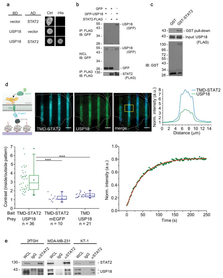
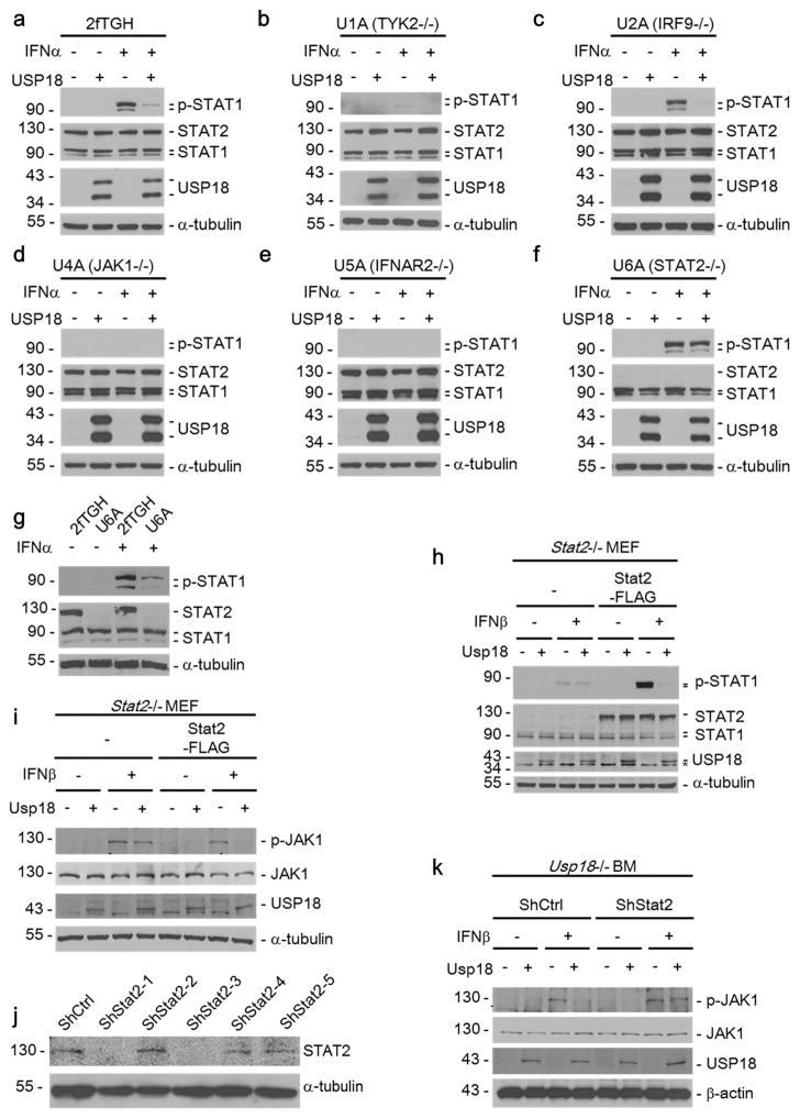
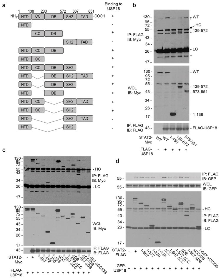
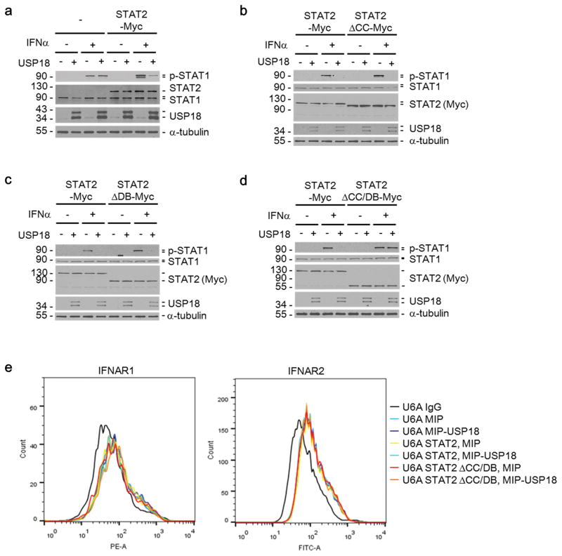
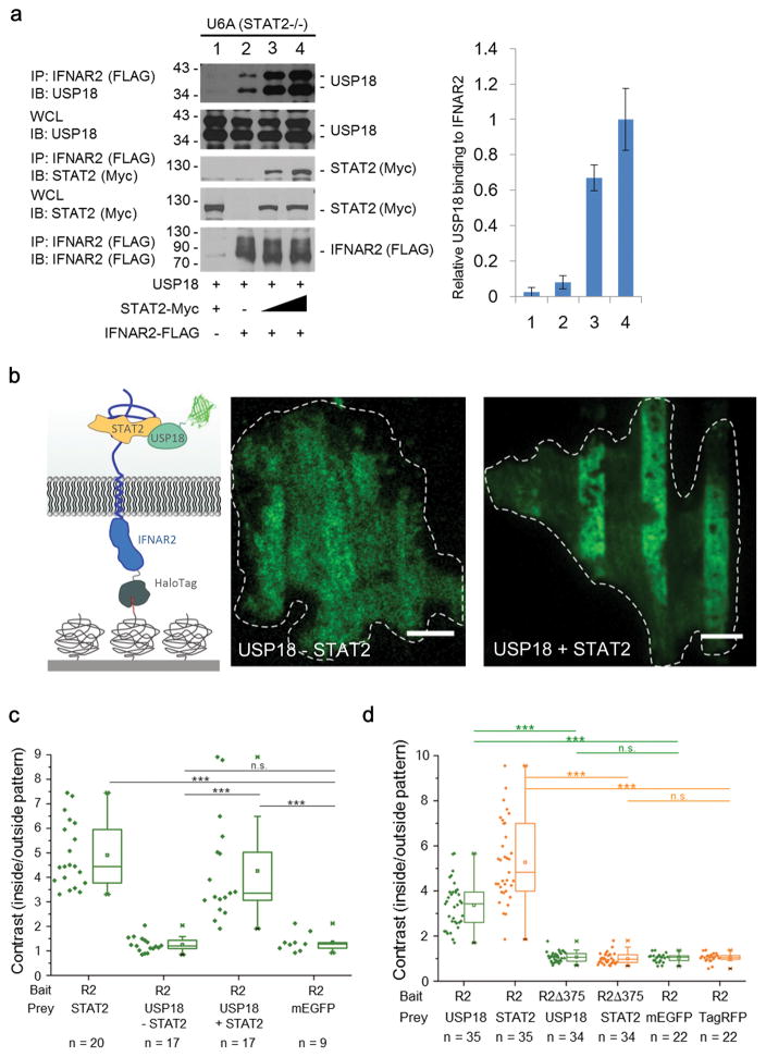
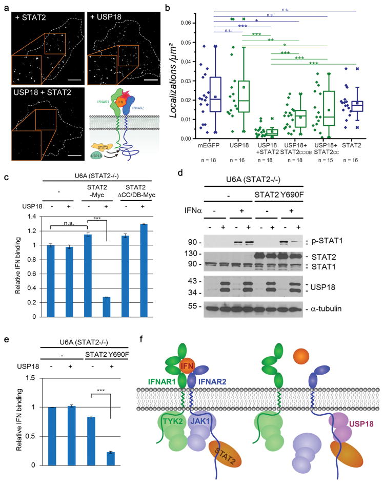
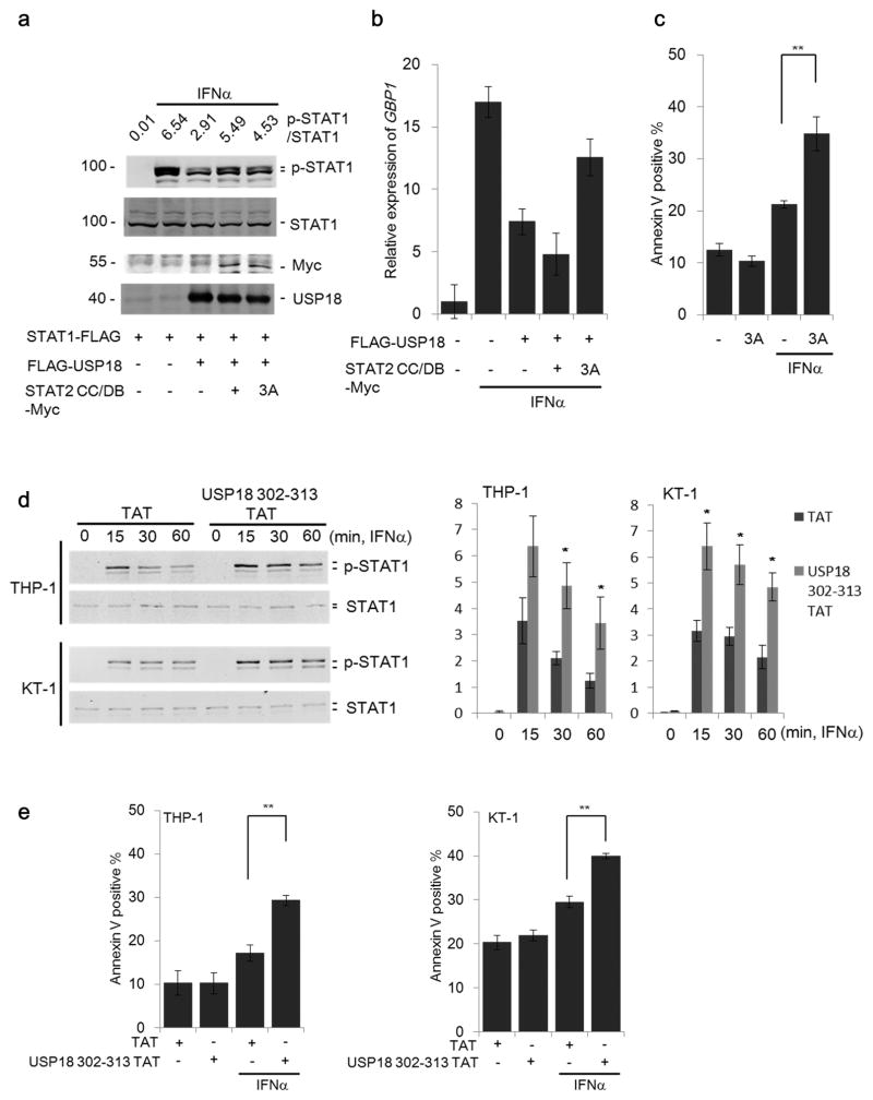
Type I interferons (IFNs) are multifunctional cytokines that regulate immune responses and cellular functions but also can have detrimental effects on human health. A tight regulatory network therefore controls IFN signaling, which in turn may interfere with medical interventions. The JAK-STAT signaling pathway transmits the IFN extracellular signal to the nucleus, thus resulting in alterations in gene expression. STAT2 is a well-known essential and specific positive effector of type I IFN signaling. Here, we report that STAT2 is also a previously unrecognized, crucial component of the USP18-mediated negative-feedback control in both human and mouse cells. We found that STAT2 recruits USP18 to the type I IFN receptor subunit IFNAR2 via its constitutive membrane-distal STAT2-binding site. This mechanistic coupling of effector and negative-feedback functions of STAT2 may provide novel strategies for treatment of IFN-signaling-related human diseases.


Conflict of interest statement

Competing financial interests

The authors declare no competing financial interests.

Figures

Figure 1
Figure 1. USP18 interacts with STAT2
(a) Yeast two-hybrid analysis of the direct interaction between USP18 and STAT2. (a),(b) Overnight cultures from strains pJ69-4a/pJ69-4alpha carrying two-hybrid plasmids with the indicated coding sequences were spotted onto selective media for plasmid maintenance (-Leu, -Tryp = Ctrl for growth control) and indicators for interactions (-His, in addition to -Leu, -Tryp). (b) Immunoblot (IB) analysis of whole cell lysates (WCL) and anti-FLAG immunoprecipitates (IP) derived from 293T cells 24 hours after co-transfection with plasmids encoding GFP, GFP-USP18, and STAT2-FLAG. The number for left side of column shows molecular weight (kDa). (c) GST pull-down assay to demonstrate that the STAT2 directly associate with USP18. (d) Immobilization of STAT2 for probing direct interaction with USP18 as depicted in the cartoon. U5A cells transfected with mEGFP-USP18 (green channel) and STAT2 intracellular fused to a transmembrane domain (TMD) and extracellular fused to mTagBFP as well as HaloTag (blue channel). Scale bars: 10 μm. Representative image of 36 cells analyzed. Intensity profiles of all channels within the yellow ROI depicted in the merged image (right panel). Quantitative analysis of the recruitment of mEGFP-USP18 to micropatterned STAT2 as determined by the contrast of the fluorescence intensities inside and outside the patterns. As negative controls, U5A cells were transfected with mEGFP-USP18 and HaloTag-mTagBFP-TMD as well as mEGFP and HaloTag-mTagBFP-TMD-STAT2. n: number of cells analyzed obtained from two independent experiments. Significance was quantified using the two-sample Kolmogorov-Smirnov test. *** P < 0.001. (lower left plot). Fluorescence recovery of USP18 recruited to micropatterned STAT2 and monoexponential fit of the recovery curve (representative of 8 cells analyzed), lower right plot). (e) Interaction of endogenous USP18 and endogenous STAT2 in 2fTGH, MDA-MB-231, and KT-1 cells. Lysates from cells treated with IFNα (1000 U/ml) for 24 hours were immunoprecipitated with IgG or anti-STAT2 and immunoblotted with STAT2 or USP18 antibodies.
Figure 2
Figure 2. STAT2 is required for USP18-mediated inhibition of Type I IFN signaling
(a–f) MIP or MIP-USP18 expressing 2fTGH, U1A, U2A, U4A, U5A, and U6A cells were treated with IFNα (1000 U/ml) for 15 minutes. The cell lysates were immunoblotted with the indicated antibodies. (g) IB analysis of STAT1 phosphorylation in 2fTGH and U6A cells in the presence or absence of (1000 U/ml) IFNα. (h–i) Stat2−/− MEFs were infected with MIP control (−) or MIP-Usp18 (+) retroviruses, either in the presence or absence of rescue with C-terminally FLAG-tagged Stat2 cDNA. Where indicated cells were treated with either mouse IFNβ (500 U/ml) for 15 minutes (h) or 30 minutes (i), before cell lysates were collected and analyzed by Western blotting with indicated antibodies. (j) Validation of Stat2 knockdown. Ba/F3 cells were infected with control or Stat2-targeting shRNA lentivirus. After 5 days puromycin selection, Stat2 expression was examined by Western blotting. (k) Usp18−/− bone marrow cells were infected with pCX4-bsr control (−) or pCX4-bsr-Usp18 (+), either in the presence or absence of control (shCtrl) or Stat2-knockdown (shStat2–3) shRNA expression. Two days following double drug selection (puromycin and blasticidin), cells were either left untreated (−) or treated (+) with mouse IFNβ (500 U/ml) for 30 minutes. Cell lysates were collected and analyzed by Western blotting with indicated antibodies.
Figure 3
Figure 3. Both the coiled-coil (CC) and DNA binding (DB) domains of STAT2 are involved in the interaction with USP18
(a) A schematic drawing of STAT2 domain structure and the associated deletion mutants used in this study. The ability of a given deletion mutant to interact with USP18 (+ or − binding) is indicated to the right. (b–c) IB analysis of WCL and anti-FLAG IP derived from 293T cells 24 hours after co-transfection with plasmids encoding FLAG-USP18 and either the full-length STAT2-Myc (WT) or the indicated deletion mutants. (d) IB analysis of WCL and anti-FLAG IP derived from 293T cells 24 hours after co-transfection with plasmids encoding GFP-USP18 and either STAT2-FLAG or the indicated deletion mutants. Asterisks are used to indicate non-specific bands. HC = Heavy Chain, LC = Light Chain.
Figure 4
Figure 4. The coiled-coil (CC) and DNA binding (DB) domains of STAT2 are important for USP18-mediated inhibition of Type I IFN signaling
(a) U6A cells, stably transduced with control (−) or C-terminally Myc-tagged STAT2 (STAT2-Myc), were infected with MIP control (−) or MIP-USP18 (+) retrovirus. With or without IFNα (1000 U/ml) treatment for 15 minutes cell lysates were collected and analyzed by Western blotting with the indicated antibodies. (b–d) U6A cells, stably transduced to express either full-length STAT2 (STAT2-Myc), or the indicated STAT2 deletion mutant (b, STAT2ΔCC-Myc; c, STAT2ΔDB-Myc; d, STAT2ΔCC/DB-Myc) with or without IFNα (1000 U/ml) treatment for 15 minutes cell lysates were collected and analyzed by Western blotting with the indicated antibodies. (e) Histograms showing the surface expression of IFNAR1 and IFNAR2 following infection with indicated constructs in U6A cell lines.
Figure 5
Figure 5. STAT2 recruits USP18 to IFNAR2
(a) IB analysis of WCL and anti-FLAG IP derived from U6A cells 24 hours after co-transfection with plasmids encoding USP18, FLAG-IFNAR2, and increasing amounts of STAT2-Myc expression construct. The relative USP18 binding to IFNAR2 from three independent experiments was quantified and plotted as the ratio of IFNAR2-bound USP18 to total USP18 (right panel). Data are normalized to the maximum binding (lane 4). (b) Recruitment of USP18 and STAT2 to micropatterned IFNAR2 in STAT2-deficient U6A cells as illustrated in the cartoon. U6A cells transfected with HaloTag-mTagBFP-IFNAR2 and mEGFP-USP18 (USP18 –STAT2) (green channel, left image) and U6A cells transfected with HaloTag-mTagBFP-IFNAR2, mEGFP-USP18 and STAT2-TagRFP-T (USP18 +STAT2) (green channel, right image). Representative images of 17 cells analyzed in two independent experiments. (c) Recruitment of USP18 to immobilized IFNAR2 (R2) into micropatterns was quantified in U6A cells by determining the contrast of the fluorescence intensities inside and outside the patterns. For comparison, constitutive binding of STAT2 (positive control) and cytosolic mEGFP (negative control) to micropatterned full length IFNAR2 expressed in U6A cells was quantified. n: number of cells analyzed in two independent experiments. Significance was quantified using the two-sample Kolmogorov-Smirnov test. *** P < 0.001, n.s., not significant. (d) The C-terminal STAT2 interacting region of IFNAR2 is necessary for recruiting USP18. Quantification of recruitment of mEGFP-USP18 co-expressed with STAT2-TagRFP-T to immobilized HaloTag-IFNAR2 or C-terminally truncated HaloTag-IFNAR2 (R2Δ375) in Hela cells. As negative controls, HeLa cells were transfected with cytosolic mEGFP or TagRFP-T, respectively. n: number of cells analyzed in two independent experiments. Significance was quantified using the two-sample Kolmogorov-Smirnov test. *** P < 0.001, n.s. not significant.
Figure 6
Figure 6. STAT2-USP18 interaction regulates ternary complex assembly of the Type I IFN receptor
(a) Binding of DY647IFNα2 M148A bound to the IFNAR at the surface of U6A cells expressing STAT2-TagRFP-T (top-left), USP18-mEGFP (top-right) or both (bottom-left). The images are superimpositions of single molecule localizations from 100 consecutive frames. Scale bars: 10 μm. Representative images of 13–18 cells analyzed for each condition. (b) Comparison of the density of DY647IFNα2 M148A bound to cell surface IFNAR of U6A cells expressing STAT2, USP18 or both proteins. Furthermore, protein with only STAT2 CC domain or with only CC and DB domains of STAT2 were also used in the assay. As a control, localizations of DY647IFNα2 M148A on the surface of U6A cells transfected with mEGFP were quantified. Data were acquired in two independent experiments, n indicates number of cells analyzed for each condition. Significance was quantified using the two-sample Kolmogorov-Smirnov test. *** P < 0.001; **P < 0.01; * P < 0.05, n.s. not significant. (c) Relative amount of FITC-labeled IFNα bound to the surface of U6A cells was examined by flow cytometry. Cells are transduced to express the indicated constructs. Data are normalized to the mean fluorescence intensity (MFI) of U6A cells in the absence of STAT2 and USP18. (d) U6A cells, stably transduced with control (−) or C-terminally Myc-tagged STAT2 Y690F mutant (STAT2 Y690F), were infected with MIP (−) or MIP-USP18 (+) retrovirus. With or without IFNα (1000 U/ml) treatment for 15 minutes, cell lysates were analyzed by Western blotting. (e) Relative amount of FITC-labeled IFNα bound to the surface of U6A cells was examined by flow cytometry. Data are normalized to the MFI of U6A cells in the absence of STAT2 and USP18. Data of figure 6 (c) and (e) are presented as mean ± S.E.M. for three independent experiments. ***P < 0.001., n.s. not significant. (f) Model of USP18-STAT2 regulating IFN response.
Figure 7
Figure 7. Inhibiting negative feedback regulation of USP18 by targeting its interaction with STAT2
(a) 293T cells were co-transfected with plasmids encoding STAT1-FLAG, FLAG-USP18 and either STAT2 CC/DB-Myc or the mutant STAT2 CC/DB L227A R409A K415A-Myc (3A). Following treatment with IFNα (1000 U/ml) for 15 minutes as indicated cell lysates were collected and immunoblotted with indicated antibodies. The ratio of p-STAT1/total STAT1 was quantified by LI-COR Odyssey system. (b) Cells indicated in Fig. 7a were treated with IFNα (1000 U/ml) for 12 hours and then expression of GBP1 was analyzed by RT q-PCR. Data represents mean ± S.D. for two independent experiments. (c) THP-1 cells transduced with either MIP control (−) or MIP-STAT2 CC/DB 3A (3A) were treated with IFNα (1000 U/ml) for 48 hours and then Annexin V positive cells were analyzed by flow cytometry. Data represents mean ± S.E.M. for three independently generated stable cell lines. **P < 0.01. (d) THP-1 and KT-1 cells were treated with TAT or USP18 aa 302-313 TAT peptide. Five hours after peptide treatment, IFNα (1000 U/ml) was added for the indicated time and cells were analyzed by Western blotting. The ratio of p-STAT1/total STAT1 from three independent experiments was quantified (right panel). Data are presented as mean +/c S.E.M. of three independent experiments. *P < 0.05. (e) TAT- or USP18 aa 302-313 TAT peptide-treated THP-1 and KT-1 cells were incubated with IFNα (1000 U/ml) for 48 hours and then Annexin V positive cells were analyzed by flow cytometry. Data are presented as mean +/− S.E.M. of three independent experiments. **P < 0.01.

Similar articles

Cited by

References

    1. Hertzog PJ, Williams BR. Fine tuning type I interferon responses. Cytokine Growth Factor Rev. 2013;24:217–25. - PubMed
    1. Schneider WM, Chevillotte MD, Rice CM. Interferon-stimulated genes: a complex web of host defenses. Annu Rev Immunol. 2014;32:513–45. - PMC - PubMed
    1. Ivashkiv LB, Donlin LT. Regulation of type I interferon responses. Nat Rev Immunol. 2014;14:36–49. - PMC - PubMed
    1. Zitvogel L, Galluzzi L, Kepp O, Smyth MJ, Kroemer G. Type I interferons in anticancer immunity. Nat Rev Immunol. 2015;15:405–14. - PubMed
    1. Borden EC, et al. Interferons at age 50: past, current and future impact on biomedicine. Nat Rev Drug Discov. 2007;6:975–90. - PMC - PubMed