. 2017 Feb 6;12(2):e0171417.
doi: 10.1371/journal.pone.0171417. eCollection 2017.

Characterization of three-dimensional cancer cell migration in mixed collagen-Matrigel scaffolds using microfluidics and image analysis

Affiliations

Characterization of three-dimensional cancer cell migration in mixed collagen-Matrigel scaffolds using microfluidics and image analysis

María Anguiano et al. PLoS One. .

Abstract

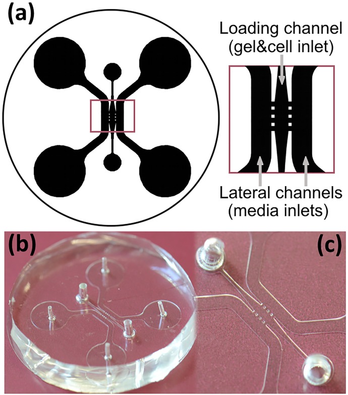
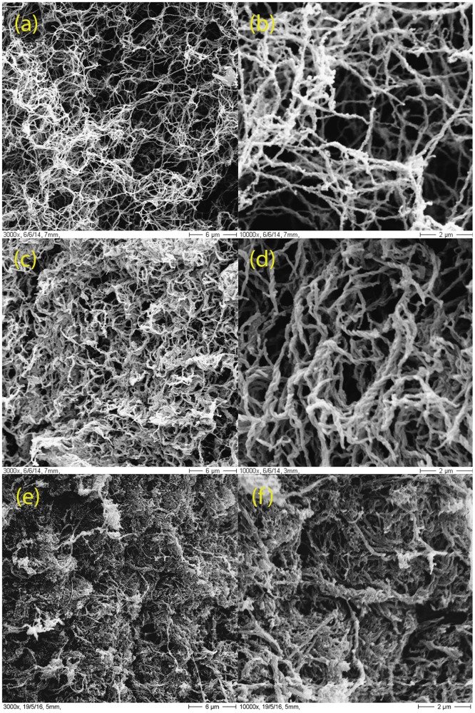
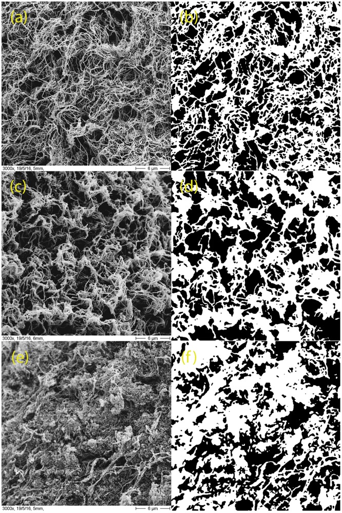
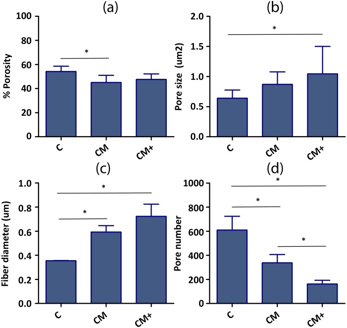
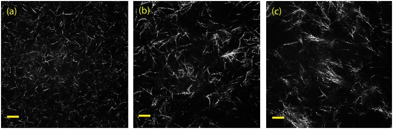
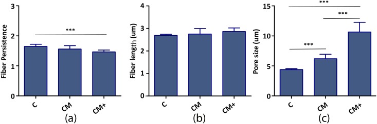
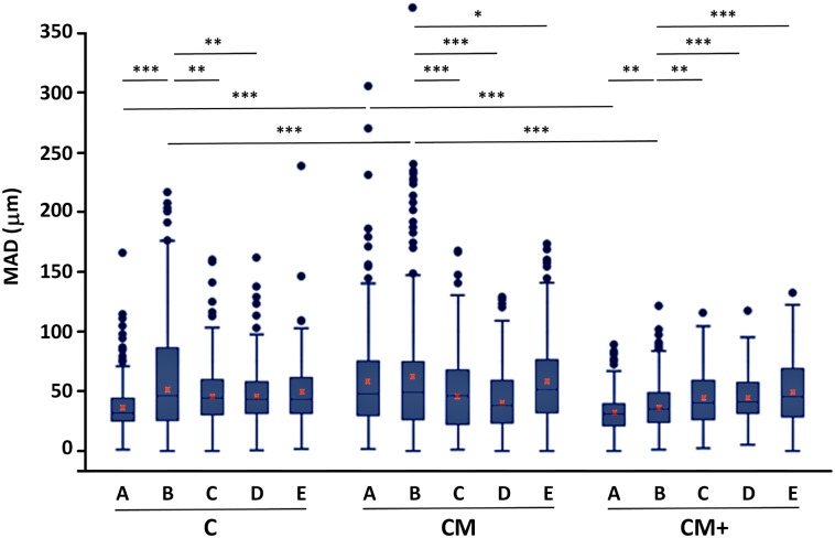
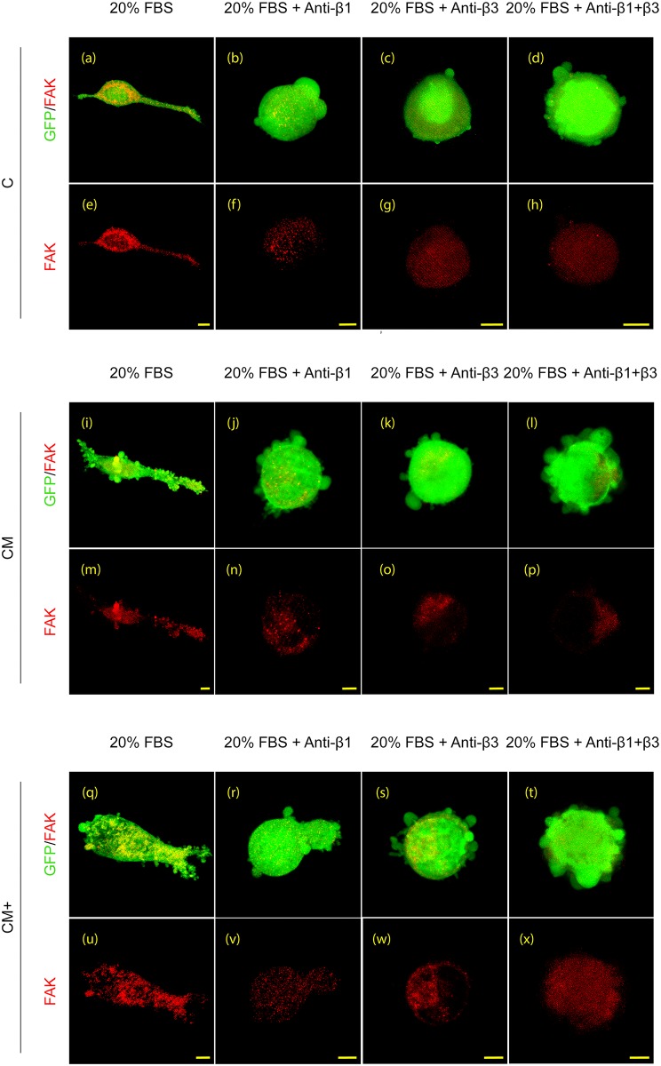
Microfluidic devices are becoming mainstream tools to recapitulate in vitro the behavior of cells and tissues. In this study, we use microfluidic devices filled with hydrogels of mixed collagen-Matrigel composition to study the migration of lung cancer cells under different cancer invasion microenvironments. We present the design of the microfluidic device, characterize the hydrogels morphologically and mechanically and use quantitative image analysis to measure the migration of H1299 lung adenocarcinoma cancer cells in different experimental conditions. Our results show the plasticity of lung cancer cell migration, which turns from mesenchymal in collagen only matrices, to lobopodial in collagen-Matrigel matrices that approximate the interface between a disrupted basement membrane and the underlying connective tissue. Our quantification of migration speed confirms a biphasic role of Matrigel. At low concentration, Matrigel facilitates migration, most probably by providing a supportive and growth factor retaining environment. At high concentration, Matrigel slows down migration, possibly due excessive attachment. Finally, we show that antibody-based integrin blockade promotes a change in migration phenotype from mesenchymal or lobopodial to amoeboid and analyze the effect of this change in migration dynamics, in regards to the structure of the matrix. In summary, we describe and characterize a robust microfluidic platform and a set of software tools that can be used to study lung cancer cell migration under different microenvironments and experimental conditions. This platform could be used in future studies, thus benefitting from the advantages introduced by microfluidic devices: precise control of the environment, excellent optical properties, parallelization for high throughput studies and efficient use of therapeutic drugs.


Conflict of interest statement

The authors have declared that no competing interests exist.

Figures

Fig 1
Fig 1. Microfluidic devices.
(a) Scheme of the device. (b) General view of a polydimethylsiloxane device. (c) Detail of the chamber that contains the hydrogel and cells (center) and the channels for medium loading (side channels).
Fig 2
Fig 2. Scanning electron microscopy (SEM).
Representative scanning electron micrographs of the three hydrogel types. (a, b) Type C; (c, d) type CM and (e,f) type CM+. The magnification shown is 3000x (left column) and 10000x (right column).
Fig 3
Fig 3. Quantification of SEM images.
Representative scanning electron micrographs of one sample image of the hydrogels and their corresponding binarized images (right column). (a, b) Type C; (c, d) type CM and (e,f) type CM+.
Fig 4
Fig 4. Quantification of hydrogel morphology from the SEM images.
(a) % Porosity, (b) Pore size area, (c) Fiber diameter, (d) Number of pores. The number of samples used to calculate %Porosity, Fiber diameter and Number of pores is three (n = 3) since we analyzed three images from each type. The number of samples used to calculate the pore size varied between sample types, since the unit used was the pore. Namely, the n values were n = 1830 (C), n = 1012 (CM) and n = 487 (CM+). * Indicates statistically significant difference of non-parametric Mann-Whitney U-test (p<0.05). Pore size dataset were compared by Anova One-Way analysis of variances followed by Bonferroni post-hoc test.
Fig 5
Fig 5. Confocal reflection microscopy.
Representative confocal reflection microscopy images of the three hydrogel types. The images are single z-slices of (a) type C; (b) type CM and (c) type CM+. The images were taken with a 63x objective. (Scale bar: 10μm).
Fig 6
Fig 6. Quantification of the morphology of hydrogels C, CM and CM+ from confocal reflection microscopy images.
(a) Fiber persistence, (b) Fiber length, (c) Pore size diameter. The number of samples used is nine (n = 9) since we analyzed nine sub-images from each type. *** indicates very statistically significant difference of Anova One-Way analysis of variances followed by Bonferroni post-hoc test (p<0.005).
Fig 7
Fig 7. Quantification of cell migration.
Box and whiskers plot of the mean accumulated distance (MAD), after 12 hours of migration hydrogel types C, CM and CM+, with no added chemo-attracting substance (A), using serum containing medium, (B) or after conjugation with integrin-blocking antibodies: 20% FBS + Anti-β1 (C), 20% FBS + Anti-β3 (D) and 20% FBS + Anti-β1+β3 (E). Red crosses mark the location of the mean value for each treatment and hydrogel, calculated as the pondered average of the mean value of the three replicas of each experiment. *, **,*** Indicates statistically significant difference with p<0.05, p<0.01 and p<0.001, respectively.
Fig 8
Fig 8. Immunophenotyping.
Representative 3D renderings of confocal immunofluorescence images showing FAK (red) staining in H1299 cells embedded in the three hydrogel types (C, CM and CM+) under the treatments described in the main text. Scale bar: 10μm.

References

    1. Chaffer CL, Weinberg RA. Perspective on cancer cell metastasis. Science. 2011; 331:1559–1564. 10.1126/science.1203543 - DOI - PubMed
    1. Fidler IJ. The pathogenesis of cancer metastasis: the 'seed and soil' hypothesis revisited. Nat Rev Cancer. 2003; 3:453–458. 10.1038/nrc1098 - DOI - PubMed
    1. Liang CC, Park AY, Guan JL. In vitro scratch assay: a convenient and inexpensive method for analysis of cell migration in vitro. Nat Protoc. 2007; 2(2):329–33. 10.1038/nprot.2007.30 - DOI - PubMed
    1. Doyle A. D., Petrie R. J., Kutys M. L., Yamada K. M. Dimensions in Cell Migration. Curr Opin in Cell Biol. 2013; 25(5), 642–649. - PMC - PubMed
    1. Shaw LM, Tumor cell invasion assays. Methods Mol Biol. 2005; 294:97–105 - PubMed

MeSH terms