. 2017 Feb 6;12(2):e0171738.
doi: 10.1371/journal.pone.0171738. eCollection 2017.

p38α regulates actin cytoskeleton and cytokinesis in hepatocytes during development and aging

Affiliations

p38α regulates actin cytoskeleton and cytokinesis in hepatocytes during development and aging

Ana M Tormos et al. PLoS One. .

Abstract

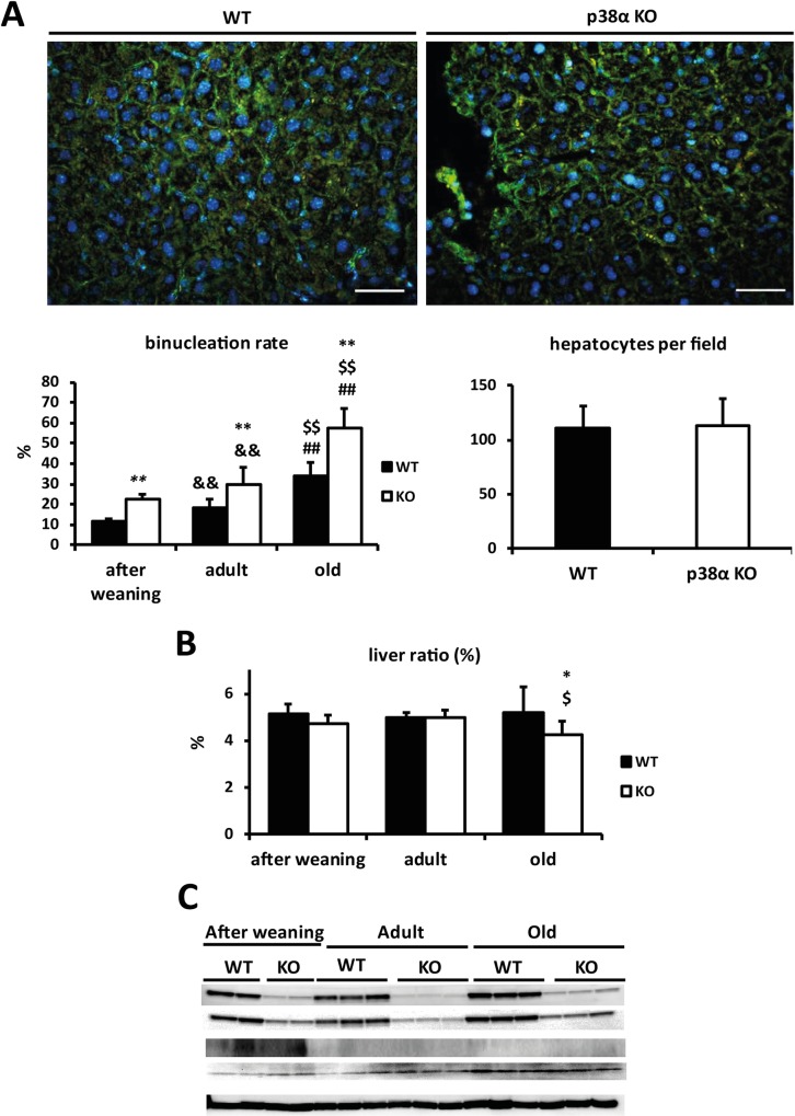
Background: Hepatocyte poliploidization is an age-dependent process, being cytokinesis failure the main mechanism of polyploid hepatocyte formation. Our aim was to study the role of p38α MAPK in the regulation of actin cytoskeleton and cytokinesis in hepatocytes during development and aging.

Methods: Wild type and p38α liver-specific knock out mice at different ages (after weaning, adults and old) were used.

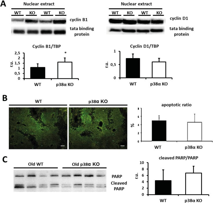
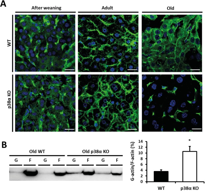
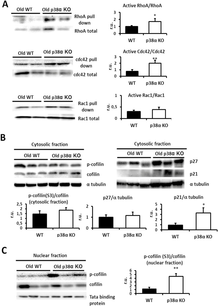
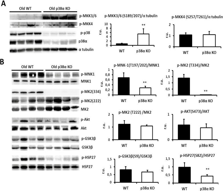
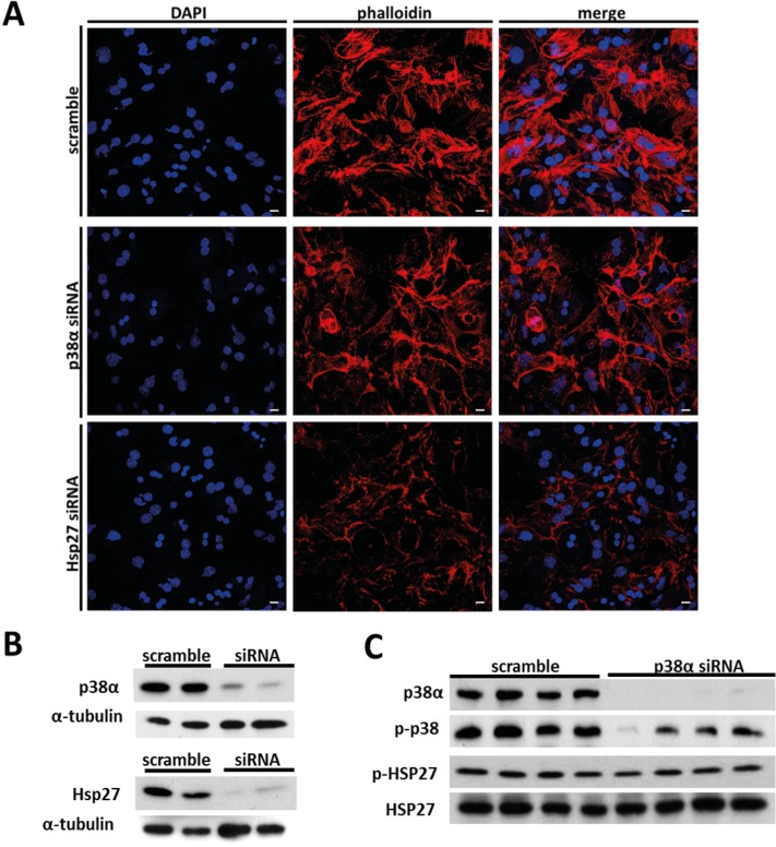
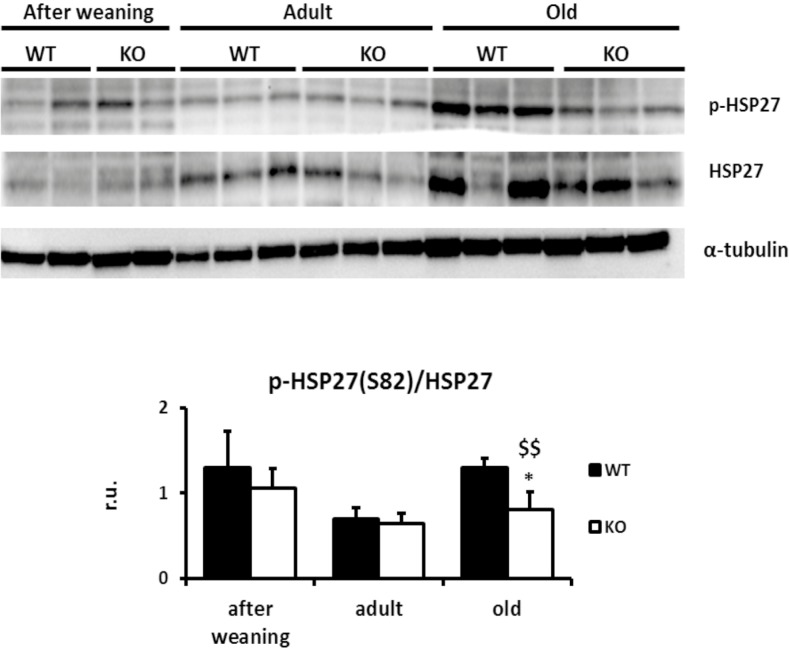
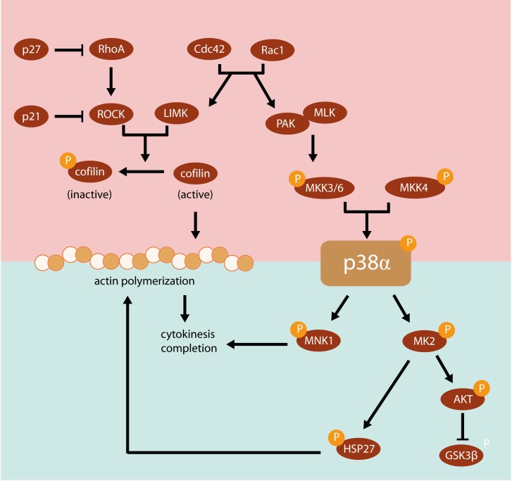
Results: We show that p38α MAPK deficiency induces actin disassembly upon aging and also cytokinesis failure leading to enhanced binucleation. Although the steady state levels of cyclin D1 in wild type and p38α knock out old livers remained unaffected, cyclin B1- a marker for G2/M transition- was significantly overexpressed in p38α knock out mice. Our findings suggest that hepatocytes do enter into S phase but they do not complete cell division upon p38α deficiency leading to cytokinesis failure and binucleation. Moreover, old liver-specific p38α MAPK knock out mice exhibited reduced F-actin polymerization and a dramatic loss of actin cytoskeleton. This was associated with abnormal hyperactivation of RhoA and Cdc42 GTPases. Long-term p38α deficiency drives to inactivation of HSP27, which seems to account for the impairment in actin cytoskeleton as Hsp27-silencing decreased the number and length of actin filaments in isolated hepatocytes.

Conclusions: p38α MAPK is essential for actin dynamics with age in hepatocytes.


Conflict of interest statement

The authors have declared that no competing interests exist.

Figures

Fig 1
Fig 1. p38α deficiency induces hepatocyte binucleation.
a. Representative image of β-catenin (green) and DAPI (blue) immunohistochemistry in old wild type and p38α knock out liver mice (Scale bars = 50 μm). Quantification of the binucleation rate (binucleated hepatocytes /total hepatocytes) with age. Number of hepatocytes per field in old wild type and p38α knock out mice as an indirect estimation of hepatocyte size in these animals. b. Liver mass ratio with age expressed as the ratio: liver weight/body weight. c. Wild type and p38α knock out livers were Western blotted for p-p38 (Thr180/Tyr182), p38α, p-H3 (Ser 10) and H3. α-tubulin was used as a loading control. Data are shown as mean ± SD. *P < 0.05, **P < 0.01 WT versus KO; &&P < 0.01 adult versus after weaning; $P < 0.05, $ $P < 0.01 old versus adult; ##P < 0.01 old versus after weaning.
Fig 2
Fig 2. p38α deficiency impairs hepatocyte cell cycle progression and does not activate pro-apoptotic pathways in old liver.
a. Nuclear fractions from old wild type and p38α knock out liver were Western blotted for cyclin B1 and cyclin D1. Tata binding protein was used as a loading control and densitometric quantification of cyclin D1/TBP and cyclin B1/TBP was performed. b. Apoptosis in wild type and p38α knock out old liver sections was measured by the number of positive nuclei using TUNEL (Scale bars = 100 μm). Apoptotic ratio was calculated as: TUNEL positive nuclei/total nuclei. c. Old wild type and p38α knock out livers were Western blotted for PARP and cleaved PARP and densitometric quantification of PARP cleavage (cleaved PARP/PARP) was performed. Data are shown as mean and SD. *P < 0.05 WT versus KO.
Fig 3
Fig 3. p38α deficiency affects actin polymerization upon aging.
a. Representative image of F-actin (green) and DAPI (blue) immunohistochemistry in after weaning, adult and old wild type and p38α knock out livers (Scale bars = 10 μm). b. F-actin and G-actin immunoblots obtained by ultracentrifugation in old wild type and p38α knock out livers. Densitometric quantification of the G-actin/F-actin ratio was performed. Data are shown as mean and SD. *P < 0.05 WT versus KO.
Fig 4
Fig 4. p38α deficiency modulates Rho GTPases.
a. Old wild type and p38α knock out livers were Western blotted for activated and total levels of RhoA, for activated and for total levels of Cdc42 and for activated and total levels of Rac1. Densitometric quantification of active RhoA/RhoA, active Cdc42/Cdc42, active Rac1/Rac1 was measured. b. Old wild type and p38α knock out liver cytosolic fractions were Western blotted for phosphorylated cofilin and total levels of cofilin, p27 and p21. α-tubulin was used as a loading control. Densitometric analysis of p-cofilin(S3)/cofilin, p27/α-tubulin and p21/α-tubulin was done. c. Old wild type and p38α knock out liver nuclear fractions were Western blotted for phosphorylated and total levels of cofilin and the densitometric quantification of p-cofilin(S3)/cofilin was performed. Tata binding protein was used as a loading control. Data are shown as mean ± SD. *P < 0.05 WT versus KO. **P < 0.01 WT versus KO.
Fig 5
Fig 5. p38α-mediated phosphorylation pathways.
a. Old wild type and p38α knock out livers were Western blotted for phosphorylated MKK3/6 and MKK4, for phosphorylated p38 and total levels of p38α. Densitometric quantification of p-MKK3/6 (S189/207)/α-tubulin, p-MKK4 (S257/T261)/a tubulin were done. b. Old wild type and p38α knock out livers were Western blotted for MNK1 and phosphorylated MNK1 on Thr197/202, for MK2 and phosphorylated MK2 on Thr334 and Thr222, for AKT and phosphorylated AKT on Ser473, for GSK3β and phosphorylated GSK3β on serine 9 and for HSP27 and phosphorylated HSP27 on Ser 82. α-tubulin was used as a loading control. Densitometric quantification of p-MNK-1(T197/202)/MNK1, p-MK2 (T334)/MK2, p-MK2 (T222) /MK2, p-AKT(S473)/AKT, p-GSK3β(S9)/GSK3β and p-HSP27(S82)/HSP27 were determined. Data are shown as mean ± SD. **P < 0.01 WT versus KO.
Fig 6
Fig 6. Actin polymerization in isolatedhepatocytesp38α MAPK-silenced and Hsp27-silenced.
a. Representative image of actin filaments staining by phalloidin (red) and DAPI (blue) in isolated hepatocytes treated with scramble, p38α MAPK siRNA and Hsp27 siRNA (Scale bars = 1000 μm). b. Silencing of p38α MAPK and Hsp27 targets by siRNA in isolated hepatocytes. c. Isolated hepatocytes scramble-treated and p38α MAPK siRNA-treated were Western blotted for p-p38 (Thr180/Tyr182), p38α, p-HSP27 (Ser82) and HSP27.
Fig 7
Fig 7. Only the in vivo animal model with a long-term p38α deficiency drives to inactivation of HSP27.
Wild type and p38α knock out livers of all groups of age were Western blotted for p-HSP27 (Ser82) and total HSP27. α-tubulin was used as a loading control. Data are shown as mean ± SD. *P < 0.05 WT versus KO; $ $P < 0.01 old versus adult.
Fig 8
Fig 8. Scheme of the p38α-mediated phosphorylation pathways involved in the regulation of actin cytoskeleton.
The Rho family plays a central role in organizing the actin cytoskeleton and in the regulation of cytokinesis. RhoA activity may be inhibited by p27, and additionally the RhoA downstream pathway may be blocked by p21 or cofilin. MNK1 and MK2 are major downstream targets of the p38α pathway that have been implicated in the regulation of cytokinesis and actin dynamics. HSP27 is another downstream target of p38α than can be also activated by MK2 and regulates the stability of actin filaments.

Similar articles

Cited by

References

    1. Gentric G, Desdouets C, Celton-Morizur S. Hepatocytes polyploidization and cell cycle control in liver physiopathology. Int J Hepatol. 2012;2012:282430 Epub 2012/11/15. 10.1155/2012/282430 - DOI - PMC - PubMed
    1. Gentric G, Celton-Morizur S, Desdouets C. Polyploidy and liver proliferation. Clin Res Hepatol Gastroenterol. 2012;36(1):29–34. Epub 2011/07/23. 10.1016/j.clinre.2011.05.011 - DOI - PubMed
    1. Gentric G, Desdouets C. Polyploidization in liver tissue. Am J Pathol. 2014;184(2):322–31. Epub 2013/10/22. 10.1016/j.ajpath.2013.06.035 - DOI - PubMed
    1. Margall-Ducos G, Celton-Morizur S, Couton D, Bregerie O, Desdouets C. Liver tetraploidization is controlled by a new process of incomplete cytokinesis. J Cell Sci. 2007;120(Pt 20):3633–9. Epub 2007/09/27. 10.1242/jcs.016907 - DOI - PubMed
    1. Celton-Morizur S, Merlen G, Couton D, Margall-Ducos G, Desdouets C. The insulin/Akt pathway controls a specific cell division program that leads to generation of binucleated tetraploid liver cells in rodents. J Clin Invest. 2009;119(7):1880–7. Epub 2009/07/16. 10.1172/JCI38677 - DOI - PMC - PubMed

MeSH terms