

Stromal alterations in ovarian cancers via wavelength dependent Second Harmonic Generation microscopy and optical scattering
- PMID: 28166758
- PMCID: PMC5294710
- DOI: 10.1186/s12885-017-3090-2
Stromal alterations in ovarian cancers via wavelength dependent Second Harmonic Generation microscopy and optical scattering
Abstract
Background: Ovarian cancer remains the most deadly gynecological cancer with a poor aggregate survival rate; however, the specific rates are highly dependent on the stage of the disease upon diagnosis. Current screening and imaging tools are insufficient to detect early lesions and are not capable of differentiating the subtypes of ovarian cancer that may benefit from specific treatments.
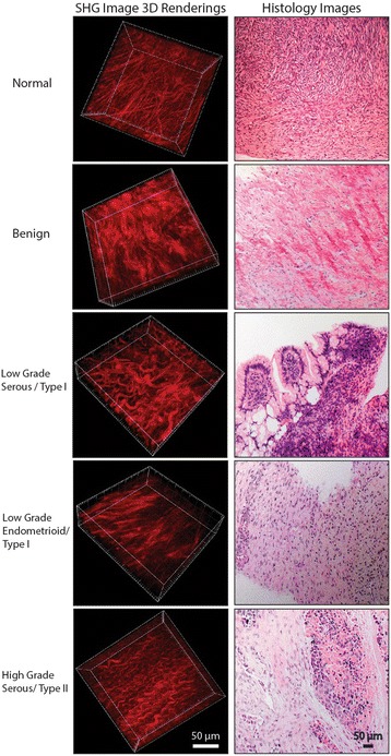
Method: As an alternative to current screening and imaging tools, we utilized wavelength dependent collagen-specific Second Harmonic Generation (SHG) imaging microscopy and optical scattering measurements to probe the structural differences in the extracellular matrix (ECM) of normal stroma, benign tumors, endometrioid tumors, and low and high-grade serous tumors.
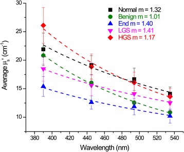
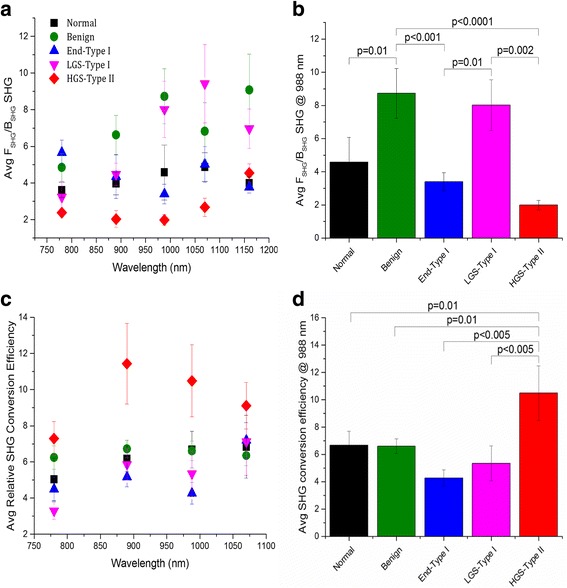
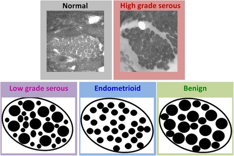
Results: The SHG signatures of the emission directionality and conversion efficiency as well as the optical scattering are related to the organization of collagen on the sub-micron size scale and encode structural information. The wavelength dependence of these readouts adds additional characterization of the size and distribution of collagen fibrils/fibers relative to the interrogating wavelengths. We found a strong wavelength dependence of these metrics that are related to significant structural differences in the collagen organization and are consistent with the dualistic classification of type I and II serous tumors. Moreover, type I endometrioid tumors have strongly differing ECM architecture than the serous malignancies. The SHG metrics and optical scattering measurements were used to form a linear discriminant model to classify the tissues, and we obtained high accuracy (>90%) between high-grade serous tumors from the other tissue types. High-grade serous tumors account for ~70% of ovarian cancers, and this delineation has potential clinical applications in terms of supplementing histological analysis, understanding the etiology, as well as development of an in vivo screening tool.
Conclusions: SHG and optical scattering measurements provide sub-resolution information and when combined provide superior diagnostic power over clinical imaging modalities. Additionally the measurements are able to delineate the different subtypes of ovarian cancer and may potentially assist in treatment protocols. Understanding the altered collagen assembly can supplement histological analysis and provide new insight into the etiology. These methods could become an in vivo screening tool for earlier detection which is important since ovarian malignancies can metastasize while undetectable by current clinical imaging resolution.
Keywords: Extracellular matrix (ECM); Optical scattering; Ovarian cancer; Second Harmonic Generation (SHG) imaging microscopy.
Figures
Similar articles
-
Polarization-resolved second harmonic generation imaging of human ovarian cancer.J Biomed Opt. 2018 Jun;23(6):1-8. doi: 10.1117/1.JBO.23.6.066501. J Biomed Opt. 2018. PMID: 29900704 Free PMC article.
-
3D texture analysis for classification of second harmonic generation images of human ovarian cancer.Sci Rep. 2016 Oct 21;6:35734. doi: 10.1038/srep35734. Sci Rep. 2016. PMID: 27767180 Free PMC article.
-
Assessing local stromal alterations in human ovarian cancer subtypes via second harmonic generation microscopy and analysis.J Biomed Opt. 2017 Nov;22(11):1-7. doi: 10.1117/1.JBO.22.11.116008. J Biomed Opt. 2017. PMID: 29188658 Free PMC article.
-
Examination of Collagen Structure and State by the Second Harmonic Generation Microscopy.Biochemistry (Mosc). 2019 Jan;84(Suppl 1):S89-S107. doi: 10.1134/S0006297919140062. Biochemistry (Mosc). 2019. PMID: 31213197 Review.
-
Precursors and pathogenesis of ovarian carcinoma.Pathology. 2013 Apr;45(3):229-42. doi: 10.1097/PAT.0b013e32835f2264. Pathology. 2013. PMID: 23478230 Review.
Cited by
-
Second-harmonic generation microscopy analysis reveals proteoglycan decorin is necessary for proper collagen organization in prostate.J Biomed Opt. 2019 May;24(6):1-8. doi: 10.1117/1.JBO.24.6.066501. J Biomed Opt. 2019. PMID: 31148435 Free PMC article.
-
Quantification of Collagen Organization after Nerve Repair.Plast Reconstr Surg Glob Open. 2017 Dec 11;5(12):e1586. doi: 10.1097/GOX.0000000000001586. eCollection 2017 Dec. Plast Reconstr Surg Glob Open. 2017. PMID: 29632766 Free PMC article.
-
Methods for the visualization and analysis of extracellular matrix protein structure and degradation.Methods Cell Biol. 2018;143:79-95. doi: 10.1016/bs.mcb.2017.08.005. Methods Cell Biol. 2018. PMID: 29310793 Free PMC article.
-
Prostate Transition Zone Fibrosis is Associated with Clinical Progression in the MTOPS Study.J Urol. 2019 Dec;202(6):1240-1247. doi: 10.1097/JU.0000000000000385. Epub 2019 Jun 12. J Urol. 2019. PMID: 31188728 Free PMC article. Clinical Trial.
-
Multiple Scattering and Random Quasi-Phase-Matching in Disordered Assemblies of LiNbO3 Nanocubes.ACS Photonics. 2022 Jun 15;9(6):1882-1888. doi: 10.1021/acsphotonics.2c00210. Epub 2022 Jun 2. ACS Photonics. 2022. PMID: 35726238 Free PMC article.
References
MeSH terms
Grants and funding
LinkOut - more resources
Full Text Sources
Other Literature Sources
Medical
Research Materials
