

Endoscopic optical coherence tomography angiography microvascular features associated with dysplasia in Barrett's esophagus (with video)
- PMID: 28167119
- PMCID: PMC5545067
- DOI: 10.1016/j.gie.2017.01.034
Endoscopic optical coherence tomography angiography microvascular features associated with dysplasia in Barrett's esophagus (with video)
Abstract
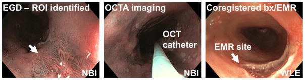
Background and aims: Angiogenesis is associated with neoplastic progression of Barrett's esophagus (BE). Volumetric optical coherence tomography angiography (OCTA) visualizes subsurface microvasculature without exogenous contrast agents. We investigated the association of OCTA microvascular features with low-grade dysplasia (LGD) and high-grade dysplasia (HGD).
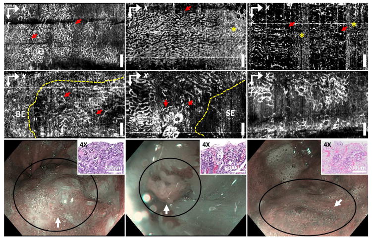
Methods: Fifty-two patients undergoing BE surveillance or endoscopic eradication therapies for dysplasia were imaged using volumetric OCTA and corresponding histologic diagnoses wre obtained to yield 97 data sets (nondysplastic BE [NDBE], 74; LGD, 10; HGD, 13). After evaluating OCTA image quality, 54 datasets (NDBE, 35; LGD, 8; HGD, 11) from 32 patients were used to develop a training and reading protocol. The association of abnormal vessel branching and heterogeneous vessel size with LGD/HGD and a regular honeycomb vessel pattern with NDBE were investigated.
Results: Blinded OCTA reading of 41 OCTA datasets (NDBE, 27; LGD, 7; HGD, 7) was performed by readers with various levels of OCT/OCTA experience including 3 OCT trainees, 1 gastroenterologist, and 2 gastroenterology fellows. Among the 6 readers, OCTA features of abnormal vessel branching and heterogeneous vessel size had an overall 94% sensitivity (95% CI, 89-99) and 69% specificity (95% CI, 62-76) for differentiating LGD/HGD versus NDBE with a mean reading time of 45 seconds per data set and moderate (kappa = .58) interobserver agreement.
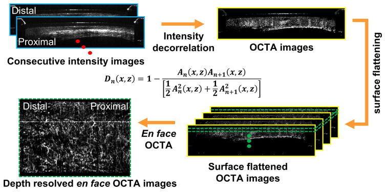
Conclusions: Volumetric en face OCTA imaging enables rapid examination of depth resolved microvascular features with near-microscopic resolution. OCTA can visualize microvascular features associated with LGD/HGD with high accuracy, which motivates new technologic advances and future studies investigating the diagnostic performance of OCTA.
Copyright © 2017 American Society for Gastrointestinal Endoscopy. Published by Elsevier Inc. All rights reserved.
Figures
References
-
- Wang KK, Sampliner RE. Updated Guidelines 2008 for the Diagnosis, Surveillance and Therapy of Barrett’s Esophagus. Am J Gastroenterol. 2008;103(3):788–797. - PubMed
-
- Devesa SS, Blot WJ, Fraumeni JF. Changing patterns in the incidence of esophageal and gastric carcinoma in the United States. Cancer. 1998;83(10):2049–2053. - PubMed
-
- Shaheen NJ. Advances in Barrett’s esophagus and esophageal adenocarcinoma. Gastroenterology. 2005;128(6):1554–1566. - PubMed
-
- Spechler SJ. Dysplasia in Barrett’s esophagus: limitations of current management strategies. Am J Gastroenterol. 2005;100(4):927–935. - PubMed
-
- Hvid-Jensen F, Pedersen L, Drewes AM, et al. Incidence of Adenocarcinoma among Patients with Barrett’s Esophagus. N Engl J Med. 2011;365(15):1375–1383. - PubMed
Publication types
MeSH terms
Grants and funding
LinkOut - more resources
Full Text Sources
Other Literature Sources
Medical
Research Materials
