Review
. 2017 Apr:37:63-70.
doi: 10.1016/j.cbpa.2017.01.013. Epub 2017 Feb 3.

Vitamin B12 in the spotlight again

Affiliations
Review

Vitamin B12 in the spotlight again

Jennifer Bridwell-Rabb et al. Curr Opin Chem Biol. 2017 Apr.

Abstract

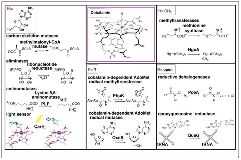
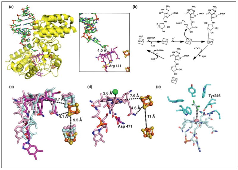
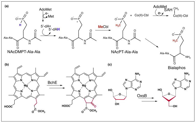
The ability of cobalamin to coordinate different upper axial ligands gives rise to a diversity of reactivity. Traditionally, adenosylcobalamin is associated with radical-based rearrangements, and methylcobalamin with methyl cation transfers. Recently, however, a new role for adenosylcobalamin has been discovered as a light sensor, and a methylcobalamin-dependent enzyme has been identified that is suggested to transfer a methyl anion. Additionally, recent studies have provided a wealth of new information about a third class of cobalamin-dependent enzymes that do not appear to use an upper ligand. They function in reductive dehalogenations and epoxide reduction reactions. Finally, mechanistic details are beginning to emerge about the cobalamin-dependent S-adenosylmethionine radical enzyme superfamily for which the role of cobalamin has been largely enigmatic.


Figures

Figure 1
Figure 1
Classes of Cbl-dependent proteins as defined by the nature of the upper ligand. AdoCbl-dependent enzymes include carbon skeleton mutases, eliminases, and aminomutases. The latter of which also requires pyridoxyl phosphate (PLP). Recently, AdoCbl has been shown to be involved in light-dependent gene regulation of the carotenoid (Car) biosynthetic operon via the transcriptional regulator CarH, adding a new function for this flavor of Cbl cofactor. MeCbl-dependent methyl transferases include methionine synthase, which catalyzes the transfer of a methyl cation to homocysteine and the mercury methylase HgcA, which is thought to catalyze transfer of a methyl anion to a Hg2+-bis(thiolate) compound (represented as Hg-S(CH3)2), the most prevalent form of Hg2+ in methylmercury producers [17]. A third class of Cbl-dependent enzymes, which includes the reductive dehalogenase PceA and the queuosine biosynthetic enzyme QueG do not have an upper axial ligand and are denoted as “open”-Cbl-dependent enzymes. The final class is made up of enzymes that appear to require the machinery for both S-adenosylmethionine (AdoMet) radical chemistry and a Cbl cofactor. Examples include phosphinate methylase PhpK and oxetanocin-A biosynthetic enzyme OxsB. The role(s) of Cbl in this enzyme family is currently under investigation.
Figure 2
Figure 2
AdoCbl functions as a light sensor in the light-dependent regulation of carotenoid biosynthesis. (a) Cartoon showing steps involved in the light-dependent regulation of the carotenoid operon by CarH. (b) Structure of the AdoCbl-bound CarH tetramer that is formed in the dark [9]. Each CarH monomer is composed of a DNA-binding domain (teal), a helical bundle (raspberry) and a Cbl-binding domain (purple) [9]. Monomers form head-to-tail dimers, and two dimers form the tetramer (other protomers in gray). AdoCbl is displayed as sticks with cyan and pink carbons for the Ado group and Cbl, respectively. (c) Structure of the CarH tetramer (colored as in panel a) bound to the carotenoid biosynthetic operon (yellow) in the dark, blocking transcription. (d) Comparison of light and dark state CarH structures. Upon exposure to sunlight, the light-sensitive Co-C bond of AdoCbl cleaves, causing a shift in the helix-bundle domain (raspberry to wheat domain movement), breaking apart the CarH dimer interface (promoter at interface in gray) and dissembling the tetramer and activating transcription [9]. Light dependent cleavage of AdoCbl results in formation of 4′,5′-anhydroadenosine [11] (cyan, boxed).
Figure 3
Figure 3
Structure and mechanism of a third class of Cbl-dependent enzymes that does not use Ado- or Me-Cbl. (a) Structure of QueG (yellow) with an oQ-modified tRNATyr anticodon stem loop bound (displayed as sticks with green carbons, left panel) [20]. The modified RNA base is inserted into a cavity in the protein, binding directly above Co of Cbl (displayed as sticks with dark pink carbons, right panel) [20]. Arg 141 (yellow carbons) of QueG blocks access to the lower axial face of Cbl. (b) The proposed mechanism of QueG. Nucleophilic attack of Co(I)-Cbl species on the oQ substrate forms an organocobalt adduct that can be converted to product and cob(II)alamin through reduction and protonation [20]. (c) An overlay of Cbls and two [4Fe-4S] clusters (orange and yellow spheres) from QueG (carbons in dark pink) and the reductive dehalogenase enzymes PceA (cyan) and NpRdhA (light pink). PceA harbors a norpseudo-B12 cofactor, which is characterized by a lower axial adenine base and a hydrogen atom at position 176 rather than a DMB base and methyl group, respectively, as found in Cbl. (d) The crystal structure of NpRdhA reveals a chloride binding site directly above Cbl [23]. Asp 471 blocks the lower face of Cbl. (e) The crystal structure of the TCE-bound PceA reveals that the distance between the substrate C and Cl atoms to Co measure 5.8- and 4.7-Å, respectively [19]. These distances do not support a direct Co-substrate interaction without invoking a conformational change. TCE is tightly packed in the active site by several bulky residues, which likely gate formation of a covalent adduct and contribute to substrate selectivity [19]. TCE is shown in two overlaid orientations as captured in the structure.
Figure 4
Figure 4
A sample of the reactions catalyzed by Cbl-dependent AdoMet radical enzymes. (a) The proposed mechanism for the Cbl-dependent AdoMet radical enzyme PhpK, which functions in the biosynthetic pathway of the commercially available herbicide bialaphos [51]. In the first step, AdoMet is reductively cleaved to generate 5′-dAdo•, which abstracts a hydrogen atom from the N-acetyldemethylphosphinothricin (NAcDMPT)-Ala-Ala substrate. The resulting radical intermediate reacts with MeCbl to form the methylated product N-acetylphosphinothricin (NAcPT)-Ala-Ala. Notably, this reaction formally involves the transfer of a methyl radical from MeCbl and regeneration of the active enzyme is thought to involve reductive methylation of Co(I)-Cbl using a second molecule of AdoMet. (b) BchE is a radical cyclase that catalyzes the conversion of magnesium protoporphyrin IX monomethyl ester to divinyl-protochlorophyllide, forming the fifth ring of bacteriochlorophyll. (c) OxsB is hypothesized to catalyze a radical mediated ring contraction of adenosine or an adenosine derivative (shown here as 2′-deoxyadenosine) in the biosynthesis of oxetanocin-A [50]. Similar to the reaction catalyzed by BchE, this reaction does not appear to require a methylation step.

Similar articles

Cited by

References

    1. Scott AI, Roessner CA. Biosynthesis of cobalamin (vitamin B(12)) Biochem Soc Trans. 2002;30:613–620. - PubMed
    1. Bassford PJ, Jr, Kadner RJ. Genetic analysis of components involved in vitamin B12 uptake in Escherichia coli. J Bacteriol. 1977;132:796–805. - PMC - PubMed
    1. Croft MT, Lawrence AD, Raux-Deery E, Warren MJ, Smith AG. Algae acquire vitamin B12 through a symbiotic relationship with bacteria. Nature. 2005;438:90–93. - PubMed
    1. Padovani D, Labunska T, Banerjee R. Energetics of interaction between the G-protein chaperone, MeaB, and B12-dependent methylmalonyl-CoA mutase. J Biol Chem. 2006;281:17838–17844. - PubMed
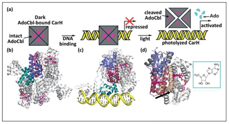
    1. Jost M, Cracan V, Hubbard PA, Banerjee R, Drennan CL. Visualization of a radical B12 enzyme with its G-protein chaperone. Proc Natl Acad Sci U S A. 2015;112:2419–2424. This work presents structural characterization of a fusion between an AdoCbl-dependent isobutyryl-CoA mutase and its G-protein chaperone. The structures presented here provide the first insight into the necessary conformational changes for loading a precious molecule of AdoCbl in an enzyme active site. - PMC - PubMed

LinkOut - more resources