. 2017 Mar 28;199(8):e00811-16.
doi: 10.1128/JB.00811-16. Print 2017 Apr 15.

A New Essential Cell Division Protein in Caulobacter crescentus

Affiliations

A New Essential Cell Division Protein in Caulobacter crescentus

Aurora Osorio et al. J Bacteriol. .

Abstract

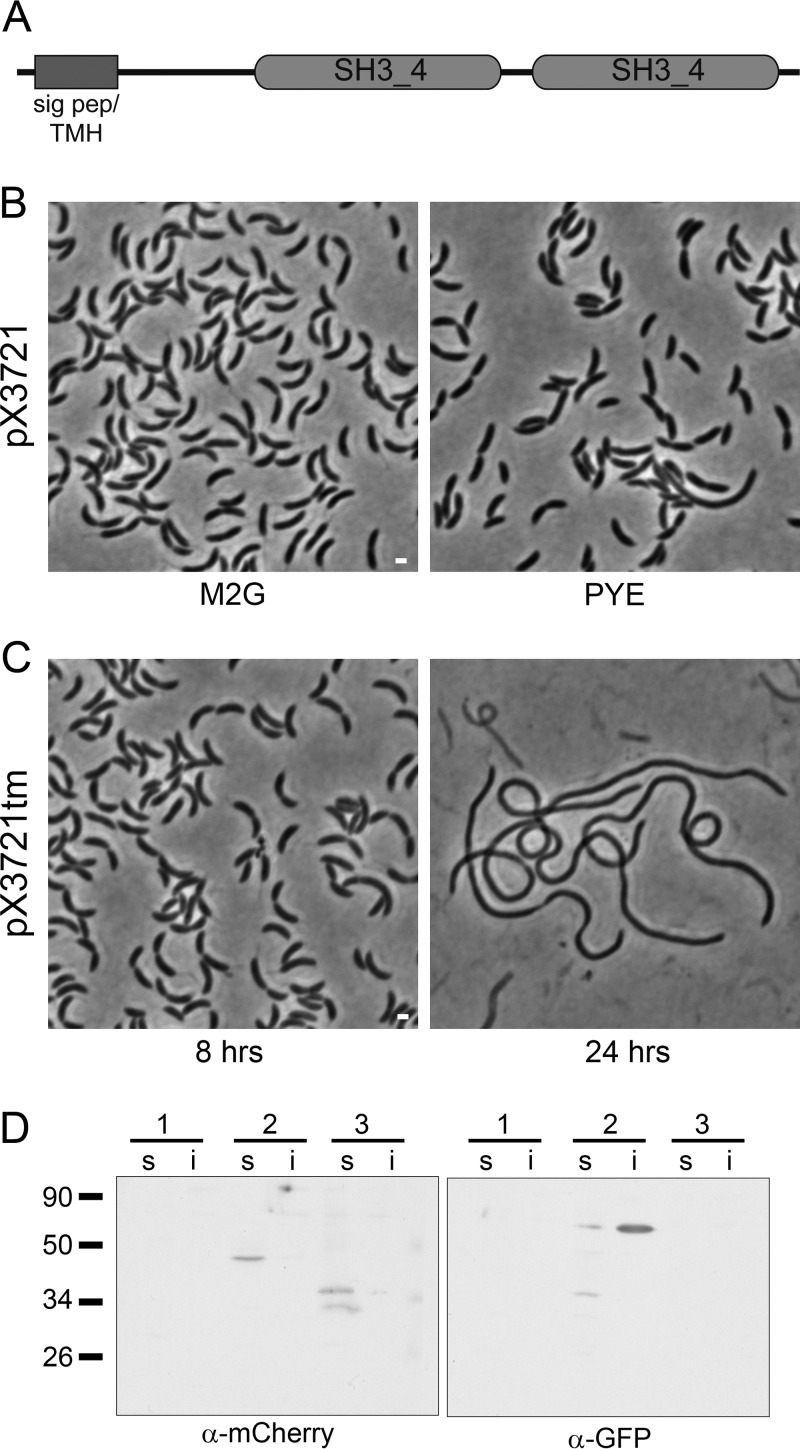
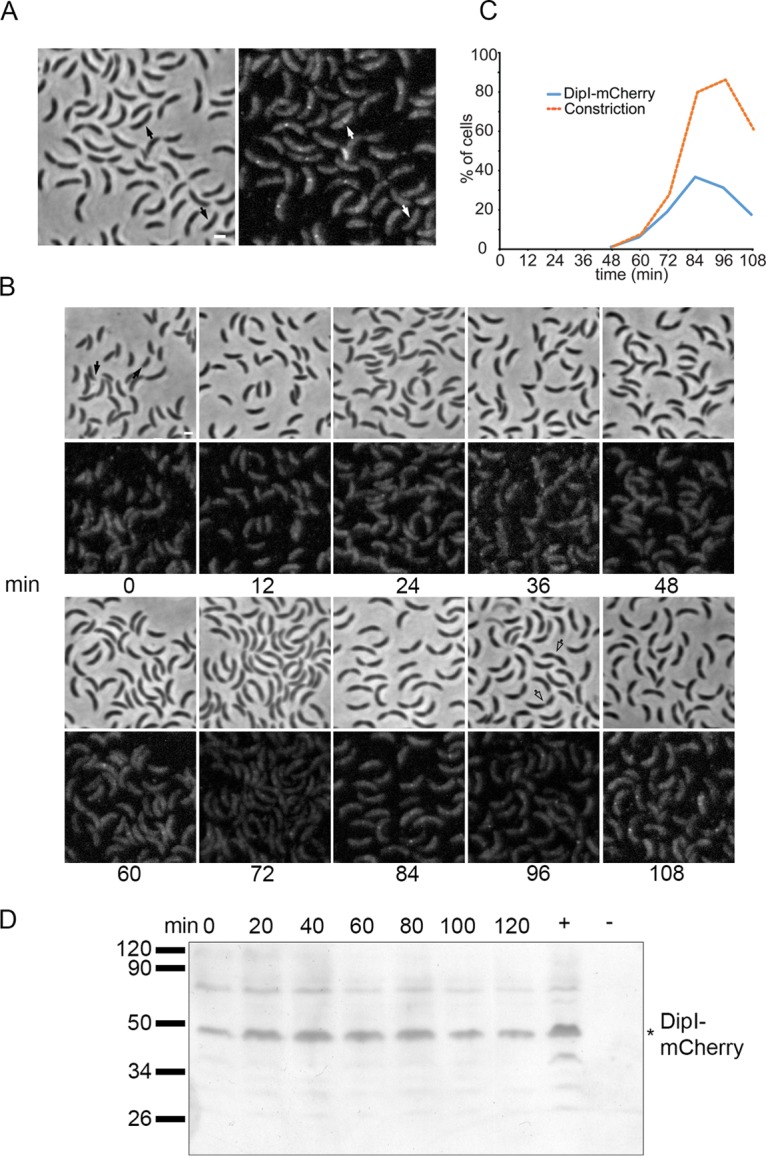
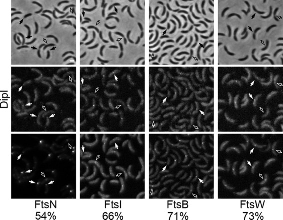
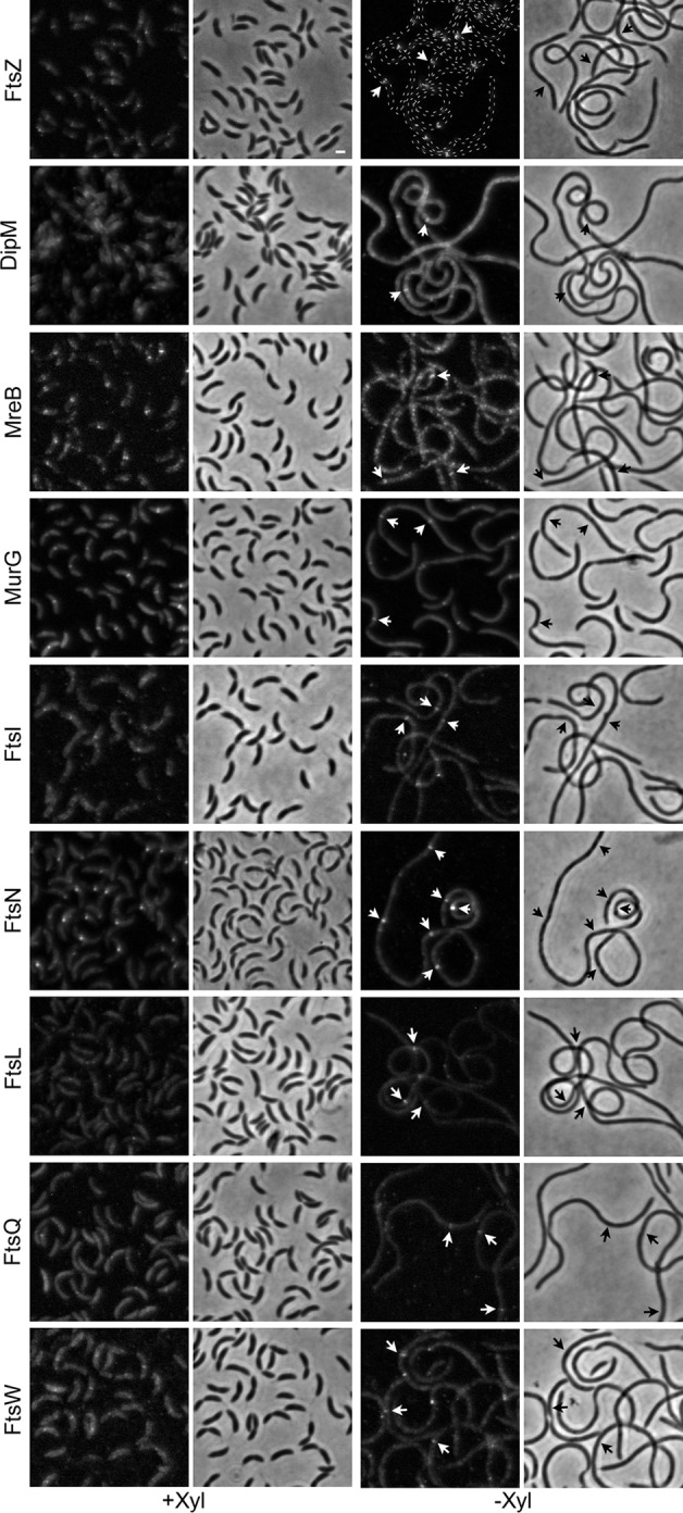
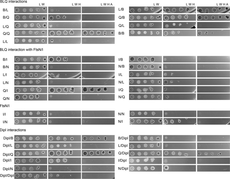
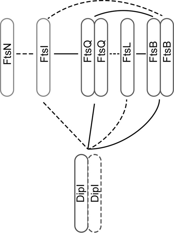
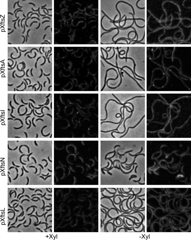
Bacterial cell division is a complex process that relies on a multiprotein complex composed of a core of widely conserved and generally essential proteins and on accessory proteins that vary in number and identity in different bacteria. The assembly of this complex and, particularly, the initiation of constriction are regulated processes that have come under intensive study. In this work, we characterize the function of DipI, a protein conserved in Alphaproteobacteria and Betaproteobacteria that is essential in Caulobacter crescentus Our results show that DipI is a periplasmic protein that is recruited late to the division site and that it is required for the initiation of constriction. The recruitment of the conserved cell division proteins is not affected by the absence of DipI, but localization of DipI to the division site occurs only after a mature divisome has formed. Yeast two-hybrid analysis showed that DipI strongly interacts with the FtsQLB complex, which has been recently implicated in regulating constriction initiation. A possible role of DipI in this process is discussed.IMPORTANCE Bacterial cell division is a complex process for which most bacterial cells assemble a multiprotein complex that consists of conserved proteins and of accessory proteins that differ among bacterial groups. In this work, we describe a new cell division protein (DipI) present only in a group of bacteria but essential in Caulobacter crescentus Cells devoid of DipI cannot constrict. Although a mature divisome is required for DipI recruitment, DipI is not needed for recruiting other division proteins. These results, together with the interaction of DipI with a protein complex that has been suggested to regulate cell wall synthesis during division, suggest that DipI may be part of the regulatory mechanism that controls constriction initiation.

Keywords: Caulobacter crescentus; SH3 domain; bacterial cell division; constriction initiation; divisome.


Figures

FIG 1
FIG 1
Depletion of DipI causes cell filamentation. (A) The domain structure of DipI is shown. (B) A strain (SP2) with a single chromosomal copy of dipI under the control of a xylose-inducible promoter (pX3721) was grown in minimal medium (M2G) or rich medium (PYE) in the absence of xylose. (C) A strain (SP3) with a single chromosomal copy of dipI fused with the sequence for the degradation signal encoded by tmRNA under the control of a xylose-inducible promoter (pX3721tm) was grown from an overnight culture in M2G medium without xylose for 8 or 24 h. (D) Presence of DipI-mCherry in the soluble fraction. Western blots of soluble (s) and insoluble (i) fractions of the following strains were probed with the indicated antibodies: 1, CB15N (wild type); 2, SP22 (expressing DipI-mCherry and Venus-FtsN protein fusions); and 3, CJW2959 (expressing periplasmic mCherry). Expected molecular masses of the proteins in kilodaltons were as follows: periplasmic mCherry, 32.8; Venus-FtsN, 55; and periplasmic DipI-mCherry, 44.2. Bars, 1 μm.
FIG 2
FIG 2
DipI is recruited late to the division site. (A) Localization of DipI-mCherry in an unsynchronized cell population. Cells from a culture of strain SP15 grown in minimal medium (M2G) were observed when the culture reached an OD660 of 0.3. Arrows indicate cells with no visible constriction in which DipI-mCherry was already localized at midcell. (B) Time-lapse images for localization of DipI-mCherry in a synchronized cell population. A culture with an OD660 of 0.3 was synchronized, and aliquots were taken for observation every 12 min. Empty arrows indicate cells with deep constriction that did not show localization of DipI. (C) Quantification of the localization of DipI to the division site. The percentage of cells that showed constriction or localization of DipI was determined in 300 cells for each time point after synchronization. (D) Presence of DipI-mCherry during the cell cycle. Total cell extracts from a synchronized culture of the SP15 strain were obtained every 15 min and the presence of DipI-mCherry was determined by Western blotting. Total cell extracts from unsynchronized SP15 and CB15N cultures were used as positive and negative controls, respectively. Migration of molecular weight markers is shown at the left. The asterisk indicates the migration of the DipI-mCherry protein. Bars, 1 μm.
FIG 3
FIG 3
DipI is recruited late to the division site. The colocalization of DipI-mCherry with other inducible cell division protein fusions was quantified in exponential cultures. Top panels, phase-contrast images; middle panels, DipI-mCherry fluorescence images; bottom panels, fluorescence images of Venus fusions with the division protein indicated at the bottom of the image (strains from left to right: SP22, SP26, SP21, and SP23). Empty arrows indicate dividing cells where localization of DipI-mCherry was expected but not observed; solid arrows indicate division sites or cell poles were colocalization was observed. The percentages of colocalization are shown at the bottom. Percentages were calculated only from cells that showed localization of the indicated division protein at the division site (n ≈ 300). Bar, 1 μm.
FIG 4
FIG 4
Mature divisomes form in the absence of DipI. The localization of different cell division proteins was determined in cells expressing DipI (+Xyl) or depleted of DipI (−Xyl). Fluorescent protein fusions of the different cell division proteins (indicated at the left of each row) were introduced as second copies and expressed from a vanillic acid-inducible promoter. The murG gene was substituted with the allele coding for the fluorescent fusion. All the strains were grown in rich medium (PYE) in the presence or absence of xylose. Depletion was carried out as described in Materials and Methods. When required, vanillic acid was added 3 h before observation. Arrows indicate localization of the division proteins in sites where no constriction was visible. From top to bottom, the strains used are as follows: SP4, SP6, SP7, SP8, SP9, SP5, SP27, SP28, and SP10. Bar, 1 μm.
FIG 5
FIG 5
DipI interacts with the FtsQLB complex. Interactions of DipI with the periplasmic domains of different cell division proteins were tested in a yeast two-hybrid assay. The mature DipI protein and the periplasmic domains of FtsQ, FtsL, FtsB, FtsI, and FtsN were fused to the activator and DNA-binding domains (AD and DBD, respectively) of the Gal4 protein, and their abilities to interact were determined by the loss of histidine auxotrophy (if the interaction was weak) and histidine and adenine auxotrophy (if it was strong). Serial dilutions of the yeast strain carrying the plasmids being tested were spotted on agar plates lacking Leu and Trp (growth control), Leu, Trp, and His (weak interaction), and Trp, Leu, His, and adenine (strong interaction). The missing amino acids or nucleotide bases are indicated at the top of each column. Protein fusions are indicated in the following order: DBD fusion/AD fusion. B, FtsB; L, FtsL; Q, FtsQ; I, FtsI; and N, FtsN.
FIG 6
FIG 6
Interaction of the cell division proteins. A summary of the results obtained in the yeast two-hybrid experiments is shown. Strong interactions (homo- or heteromeric) are shown as solid lines and weak interactions are shown as broken lines.
FIG 7
FIG 7
Maturation of the divisome is required for DipI recruitment. The localization of DipI-mCherry was determined in cells depleted of different cell division proteins (indicated at the left of each row). Cells were grown in PYE and depletion was carried out as described in Materials and Methods. Strains used from top to bottom are as follows: SP29, SP16, SP18, SP19, and SP17. Bar, 1 μm.

References

    1. Nanninga N. 1991. Cell division and peptidoglycan assembly in Escherichia coli. Mol Microbiol 5:791–795. doi:10.1111/j.1365-2958.1991.tb00751.x. - DOI - PubMed
    1. Egan AJ, Vollmer W. 2013. The physiology of bacterial cell division. Ann N Y Acad Sci 1277:8–28. doi:10.1111/j.1749-6632.2012.06818.x. - DOI - PubMed
    1. Vicente M, Rico AI, Martinez-Arteaga R, Mingorance J. 2006. Septum enlightenment: assembly of bacterial division proteins. J Bacteriol 188:19–27. doi:10.1128/JB.188.1.19-27.2006. - DOI - PMC - PubMed
    1. Moll A, Thanbichler M. 2009. FtsN-like proteins are conserved components of the cell division machinery in proteobacteria. Mol Microbiol 72:1037–1053. doi:10.1111/j.1365-2958.2009.06706.x. - DOI - PubMed
    1. Bi EF, Lutkenhaus J. 1991. FtsZ ring structure associated with division in Escherichia coli. Nature 354:161–164. doi:10.1038/354161a0. - DOI - PubMed

Publication types

MeSH terms

Substances

LinkOut - more resources