Comparative Study
. 2017 Mar 10;292(10):4326-4335.
doi: 10.1074/jbc.M117.779868. Epub 2017 Feb 6.

Adenosine Deaminase That Acts on RNA 3 (ADAR3) Binding to Glutamate Receptor Subunit B Pre-mRNA Inhibits RNA Editing in Glioblastoma

Affiliations
Comparative Study

Adenosine Deaminase That Acts on RNA 3 (ADAR3) Binding to Glutamate Receptor Subunit B Pre-mRNA Inhibits RNA Editing in Glioblastoma

Eimile Oakes et al. J Biol Chem. .

Abstract

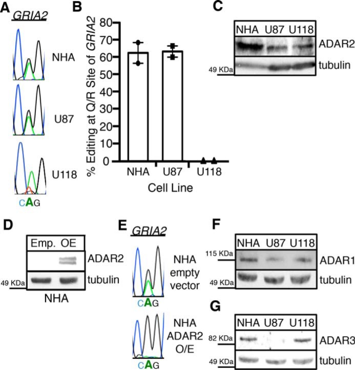
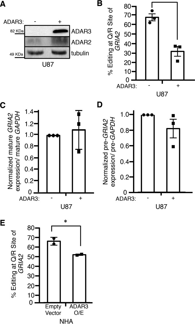
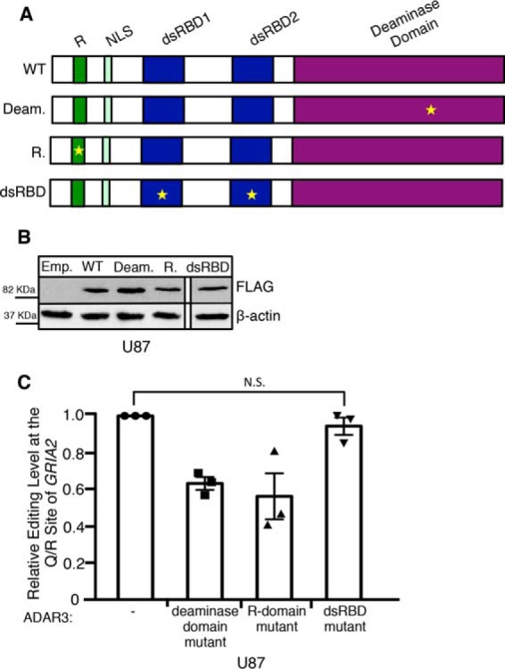
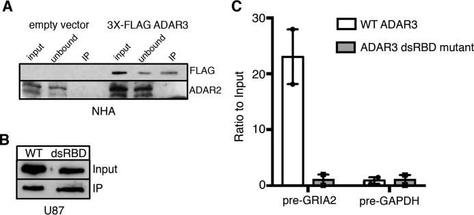
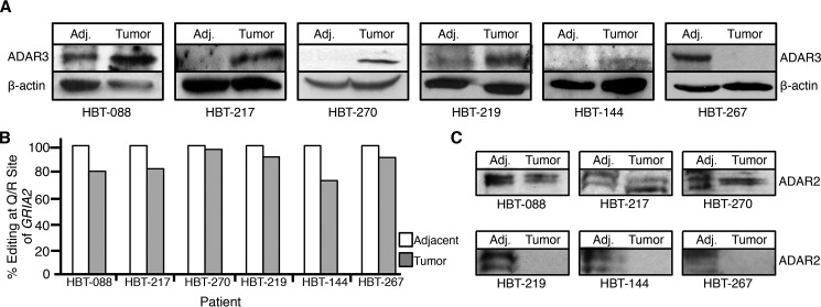
RNA editing is a cellular process that precisely alters nucleotide sequences, thus regulating gene expression and generating protein diversity. Over 60% of human transcripts undergo adenosine to inosine RNA editing, and editing is required for normal development and proper neuronal function of animals. Editing of one adenosine in the transcript encoding the glutamate receptor subunit B, glutamate receptor ionotropic AMPA 2 (GRIA2), modifies a codon, replacing the genomically encoded glutamine (Q) with arginine (R); thus this editing site is referred to as the Q/R site. Editing at the Q/R site of GRIA2 is essential, and reduced editing of GRIA2 transcripts has been observed in patients suffering from glioblastoma. In glioblastoma, incorporation of unedited GRIA2 subunits leads to a calcium-permeable glutamate receptor, which can promote cell migration and tumor invasion. In this study, we identify adenosine deaminase that acts on RNA 3 (ADAR3) as an important regulator of Q/R site editing, investigate its mode of action, and detect elevated ADAR3 expression in glioblastoma tumors compared with adjacent brain tissue. Overexpression of ADAR3 in astrocyte and astrocytoma cell lines inhibits RNA editing at the Q/R site of GRIA2 Furthermore, the double-stranded RNA binding domains of ADAR3 are required for repression of RNA editing. As the Q/R site of GRIA2 is specifically edited by ADAR2, we suggest that ADAR3 directly competes with ADAR2 for binding to GRIA2 transcript, inhibiting RNA editing, as evidenced by the direct binding of ADAR3 to the GRIA2 pre-mRNA. Finally, we provide evidence that both ADAR2 and ADAR3 expression contributes to the relative level of GRIA2 editing in tumors from patients suffering from glioblastoma.

Keywords: ADAR; ADAR3; GRIA2; RNA editing; adenosine deaminase; cancer; double-stranded RNA (dsRNA); glioblastoma; glutamate receptor; inosine.


Conflict of interest statement

The authors declare that they have no conflicts of interest with the contents of this article

Figures

FIGURE 1.
FIGURE 1.
ADAR3 expression is consistent with an inhibitor of GRIA2 editing in astrocyte- and grade IV astrocytoma-derived cell lines. A, chromatograms of the Q/R site of GRIA2 in NHA, U87, and U118 cells. Black indicates transcripts with guanosine (edited), and green indicates transcripts with adenosine (unedited). B, editing levels at the Q/R site of GRIA2 were measured in the indicated cell lines (n = 2). Error bars represent S.E. C, equivalent amounts of cell lysates were determined by Bradford assay and subjected to SDS-PAGE and immunoblotting for ADAR2. Tubulin is the loading control. D, lysates from NHA cells transduced with retrovirus containing a neomycin resistance vector with no protein (Emp.) or human ADAR2 expressed from the CMV promoter (OE) were subjected to SDS-PAGE and immunoblotted for ADAR2. Tubulin is the loading control. E, chromatograms of the Q/R site of GRIA2 in the cell lines show in D. Black indicates transcripts with guanosine (edited), and green indicates transcripts with adenosine (unedited). Bold A indicates the genomically encoded adenosine at the Q/R site of GRIA2. F and G, equivalent amounts of cell lysates were determined by Bradford assay and subjected to SDS-PAGE and immunoblotting for ADAR1 (F) or ADAR3 (G). Tubulin is the loading control.
FIGURE 2.
FIGURE 2.
ADAR3 inhibits RNA editing at the Q/R site of GRIA2. A, equivalent amounts of lysates from U87 cells transduced with a retrovirus containing a neomycin resistance vector with no protein (−) or human ADAR3 expressed from the CMV promoter (+) were determined by Bradford assay and subjected to SDS-PAGE and immunoblotting for ADAR2 and ADAR3. Tubulin antibody was used as a loading control. B, editing levels at the Q/R site of GRIA2 were measured in U87 cells described in A for three independent biological replicates. Error bars represent S.E., and a significant (p value <0.005) change in editing level is denoted by the asterisk. C and D, the levels of mature (C) and precursor (D) GRIA2 mRNA and control GAPDH mRNA were determined by qRT-PCR for three independent biological replicates. The bar height represents the relative ratio of GRIA2 transcript to GAPDH transcripts normalized to the value obtained for the U87 cells transduced with a retrovirus containing a neomycin resistance vector with no protein (−). Error bars represent S.E. E, editing levels at the Q/R site of GRIA2 were measured in NHA cells transduced with retrovirus containing a neomycin resistance vector with no protein (Empty Vector) or human ADAR3 expressed from the CMV promoter (ADAR3 O/E) for two independent biological replicates. Error bars represent S.E., and a significant (p value <0.05) change in editing level is denoted by the asterisk.
FIGURE 3.
FIGURE 3.
The dsRBDs of ADAR3 are required for inhibition of editing at the Q/R site of GRIA2. A, schematic of wild-type ADAR3 and ADAR3 with mutations in annotated domains. Regions annotated are the R-domain (R), nuclear localization signal (NLS), dsRBD1 and dsRBD2, and deaminase domain. Stars indicate the location of point mutation(s) in each mutant construct. B, Western blot of lysates from U87 cells expressing an integrated neomycin resistance vector with no protein (emp.) or expressing CMV-driven human wild-type ADAR3 (WT), ADAR3 with a point mutation to the deaminase domain (Deam.), ADAR3 with mutations to the R-domain (R.), or ADAR3 with mutations to the dsRBDs (dsRBD mutant lysate was run together on the same gel, but one lane is cropped out) subjected to SDS-PAGE and immunoblotting. All ADAR3 expression constructs contain an N-terminal FLAG tag and were detected with FLAG antibody. Actin is the loading control. C, editing levels at the Q/R site of GRIA2 were measured in the indicated cell lines for three independent biological replicates. The bar height represents the percentage of GRIA2 transcripts edited in each cell line normalized to U87 cells expressing a neomycin resistance vector with no protein (−). Error bars represent S.E. N.S. is not significant (p > 0.3), calculated using a two-tailed t test.
FIGURE 4.
FIGURE 4.
ADAR3 binds directly to GRIA2 transcripts. A, lysates from NHA cells transduced with a retrovirus containing a neomycin resistance vector with no protein (empty vector) or 3X-FLAG-tagged human ADAR3 expressed from the CMV promoter were subjected to incubation with α-FLAG magnetic beads. Input represents lysates before incubation with beads (0.5%, 3%:FLAG, ADAR2 blots), unbound represents lysates after immunoprecipitation (0.5%, 3%:FLAG, ADAR2 blots), and IP represents protein bound to FLAG beads (10%, 50%:FLAG, ADAR2 blots). Immunoblotting for the FLAG epitope (ADAR3) and ADAR2 was performed. B, Western blot of input lysates and immunoprecipitation lysates from 3X-FLAG-ADAR3 (WT) and 3X-FLAG-ADAR3 with mutations to the dsRBD. C, after treating immunoprecipitates from B with proteinase K, RNA was extracted and reverse transcribed with gene-specific primers. Bar heights indicate the ratio of cDNA in the immunoprecipitates over the inputs relative to input cDNA in U87 cells expressing wild-type ADAR3 and ADAR3 with mutations to the dsRNA binding domains normalized to the average of the two experiments (n = 2; error bars represent S.E.).
FIGURE 5.
FIGURE 5.
ADAR3 expression is elevated in a majority of glioblastoma patient tumors. A, lysates from glioblastoma tissue (Tumor) and matched adjacent brain tissue (Adj.) were subjected to SDS-PAGE and immunoblotting for ADAR3. Actin is the loading control. B, percentage of mRNAs edited at the Q/R site of GRIA2 in glioblastoma tissue and matched adjacent brain tissue from six glioblastoma patients. C, lysates from glioblastoma tissue (Tumor) and matched adjacent brain tissue (Adj.) were subjected to SDS-PAGE and immunoblotting for ADAR2.

References

    1. Nishikura K. (2010) Functions and regulation of RNA editing by ADAR deaminases. Annu. Rev. Biochem. 79, 321–349 - PMC - PubMed
    1. Yang Y., Zhou X., and Jin Y. (2013) ADAR-mediated RNA editing in non-coding RNA sequences. Sci. China Life Sci. 56, 944–952 - PubMed
    1. Savva Y. A., Rieder L. E., and Reenan R. A. (2012) The ADAR protein family. Genome Biol. 13, 252. - PMC - PubMed
    1. Bass B. L. (2002) RNA editing by adenosine deaminases that act on RNA. Annu. Rev. Biochem. 71, 817–846 - PMC - PubMed
    1. Pullirsch D., and Jantsch M. F. (2010) Proteome diversification by adenosine to inosine RNA editing. RNA Biol. 7, 205–212 - PubMed

Publication types

MeSH terms

LinkOut - more resources