. 2017 Mar 31;292(13):5429-5442.
doi: 10.1074/jbc.M116.770131. Epub 2017 Feb 6.

Adaptation of a Genetic Screen Reveals an Inhibitor for Mitochondrial Protein Import Component Tim44

Affiliations

Adaptation of a Genetic Screen Reveals an Inhibitor for Mitochondrial Protein Import Component Tim44

Non Miyata et al. J Biol Chem. .

Abstract

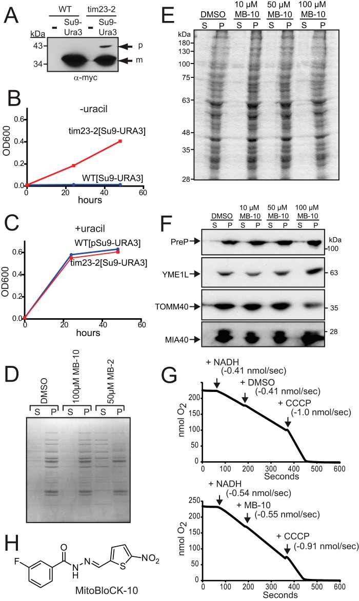
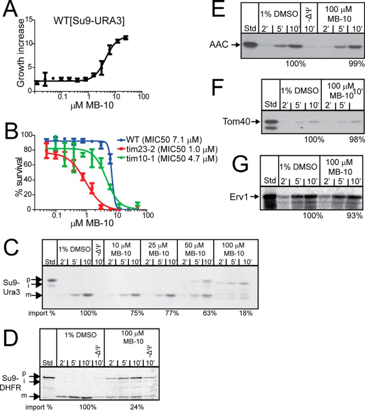
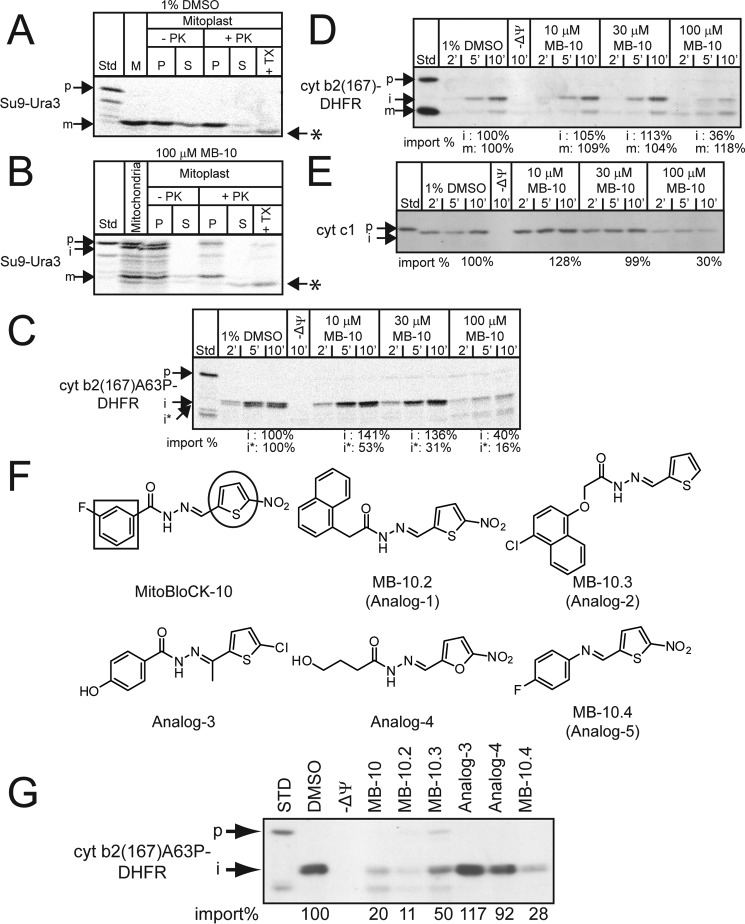
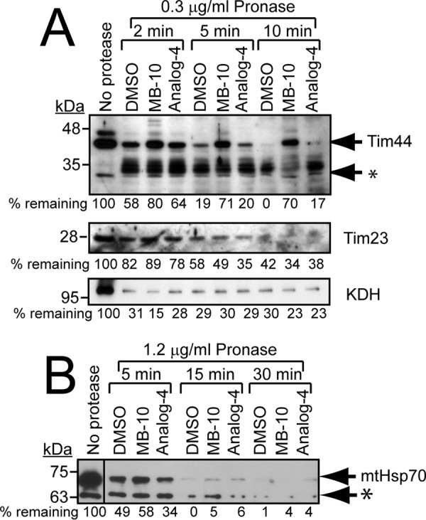
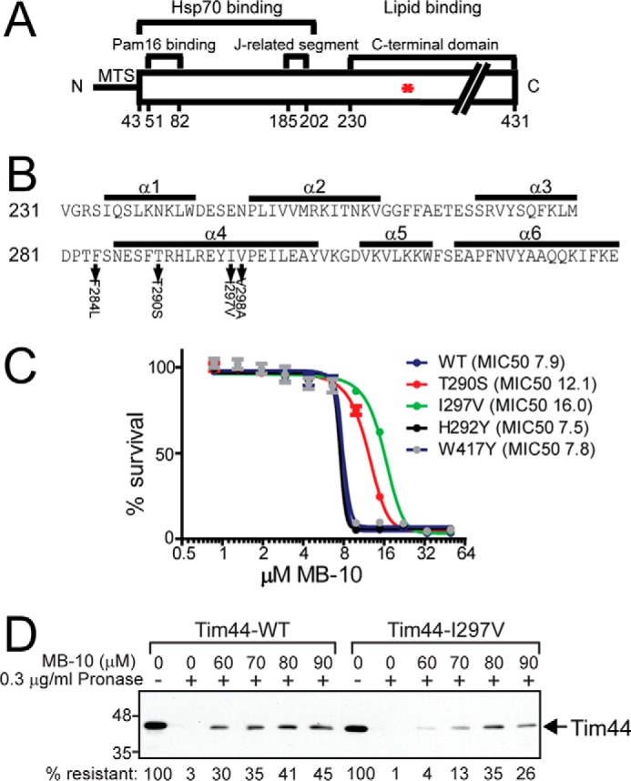
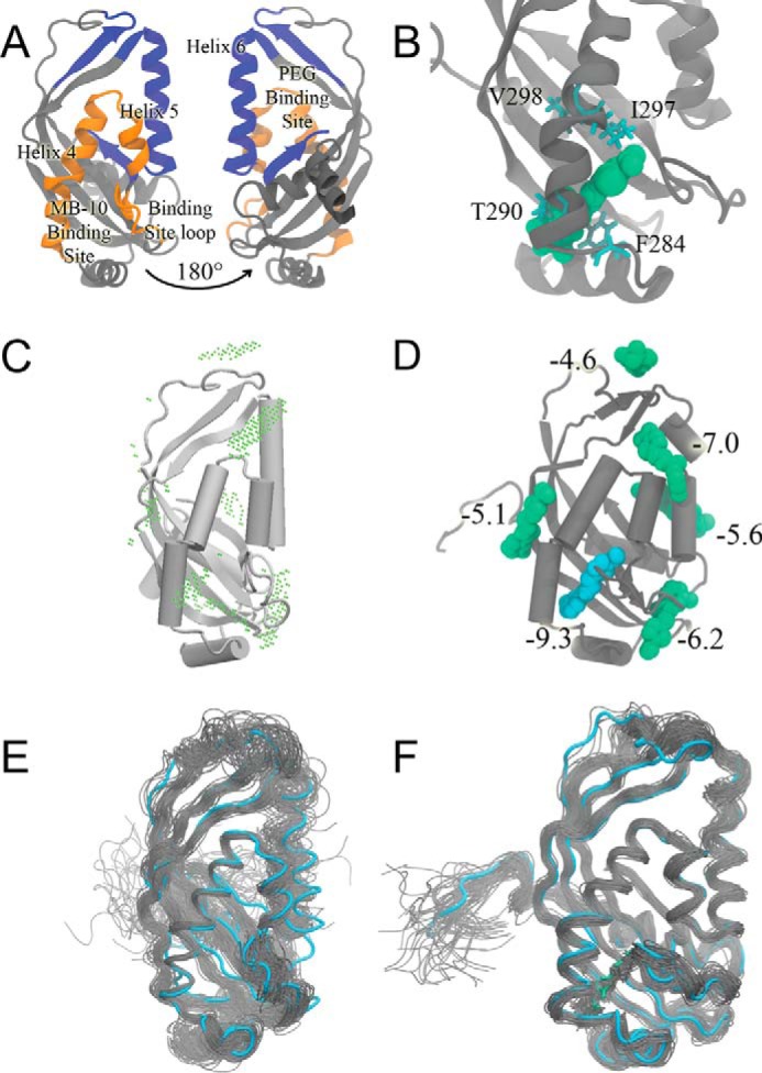
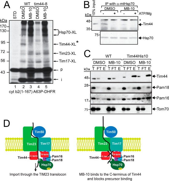
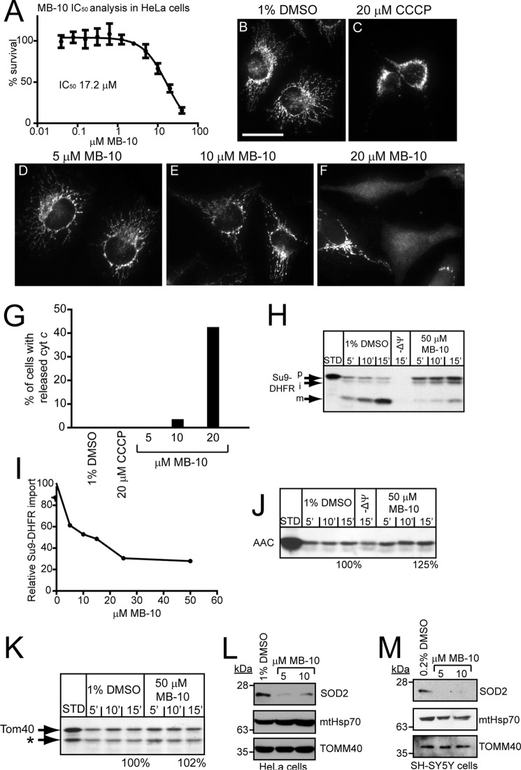
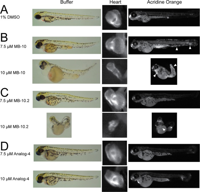
Diverse protein import pathways into mitochondria use translocons on the outer membrane (TOM) and inner membrane (TIM). We adapted a genetic screen, based on Ura3 mistargeting from mitochondria to the cytosol, to identify small molecules that attenuated protein import. Small molecule mitochondrial import blockers of the Carla Koehler laboratory (MB)-10 inhibited import of substrates that require the TIM23 translocon. Mutational analysis coupled with molecular docking and molecular dynamics modeling revealed that MB-10 binds to a specific pocket in the C-terminal domain of Tim44 of the protein-associated motor (PAM) complex. This region was proposed to anchor Tim44 to the membrane, but biochemical studies with MB-10 show that this region is required for binding to the translocating precursor and binding to mtHsp70 in low ATP conditions. This study also supports a direct role for the PAM complex in the import of substrates that are laterally sorted to the inner membrane, as well as the mitochondrial matrix. Thus, MB-10 is the first small molecule modulator to attenuate PAM complex activity, likely through binding to the C-terminal region of Tim44.

Keywords: chemical biology; mitochondria; mitochondrial transport; protein translocation; small molecule.


Conflict of interest statement

The authors declare that they have no conflicts of interest with the contents of this article

Figures

FIGURE 1.
FIGURE 1.
MB-10 is a potential attenuator of protein import into mitochondria. A, a plasmid expressing Su9-Ura3-myc was integrated at the LEU2 locus in WT and tim23-2 strains, and the strains were grown at 30 °C. A whole cell lysate from WT and the tim23-2 mutant, either without (−) or with [Su9-Ura3], was separated by SDS-PAGE followed by immunoblotting with an anti-Myc antibody. p, precursor; m, mature. B, growth analysis of the WT and tim23-2 mutant with integrated [Su9-URA3] in synthetic dextrose medium in the absence of uracil. Growth was measured based on the density at A600 (OD600). C, as in B, in the presence of uracil. D, 1% DMSO, 100 μm MB-10, or 50 μm MB-2 was added to purified 100 μg/ml WT yeast mitochondria for 30 min at 25 °C in import buffer, and released proteins (S) were separated from mitochondria (P) by centrifugation. The proteins were visualized by Coomassie staining. E, as in B, 1% DMSO or the indicated concentration of MB-10 was added to purified mitochondria from HEK293T cells followed by Coomassie staining. F, as in E, the release of proteins was detected by immunoblotting; proteins included PreP (matrix), YME1L (inner membrane), TOMM40 (outer membrane), and MIA40 (intermembrane space). G, respiration measurements were performed with a Clark-type oxygen electrode using 100 μg/ml WT yeast mitochondria in the presence of 1% DMSO or MB-10. Respiration was initiated with NADH addition. 100 μm MB-10 (with DMSO at a 1% final concentration) or 1% DMSO was added once steady-state respiration was established. As a control, CCCP was added to uncouple the mitochondria. The respiration rate for each treatment has been included. H, the structure of MitoBloCK-10 (MB-10).
FIGURE 2.
FIGURE 2.
MB-10 inhibits the import of substrates that use the TIM23 import pathway. A, the WT yeast strain expressing Su9-Ura3 was cultured in synthetic dextrose medium lacking uracil in the indicated concentrations of MB-10 for 24 h. The cell density is indicated as fold increase compared with growth in the presence of DMSO. The data represent the averages ± S.D. of n = 3 trials. B, MIC50 analysis of WT, tim23-2, and tim10-1 strains with MB-10. Each strain was cultured in rich ethanol-glycerol medium in the presence of various concentrations of MB-10 for 24 h. 100% was set as A600 of each strain grown in the presence of the vehicle control (1% DMSO). Average percentage of survival ± S.D. of n = 3 trials is shown. C–G, import assays were performed with radiolabeled precursors into mitochondria from the WT strain in the presence of MB-10 or the vehicle (1% DMSO). Non-imported precursor was removed by trypsin treatment. Precursors include Su9-Ura3 (C), Su9-DHFR (D), AAC (E), Tom40 (F), and Erv1 (G). 10% standard (Std) from the translation reaction is included. A representative gel is shown for each assay (n = 3). The last time point in the import reactions was quantified with ImageJ software; 100% was set as the amount of precursor imported in the presence of DMSO at end point in the time course. p, precursor; m, mature; i, intermediate.
FIGURE 3.
FIGURE 3.
MB-10 inhibition of import depends on specific chemical characteristics. A and B, Su9-Ura3 was imported as in Fig. 2C. After import in the presence of 1% DMSO (A), or 100 μm MB-10 (B), samples were incubated in hypotonic buffer to swell the outer membrane (generating mitoplasts) in the presence and absence of proteinase K (PK) followed by inactivation with PMSF. Mitoplasts were recovered by centrifugation (P) and separated from supernatant (S), which contains the soluble intermembrane space contents. As a control, samples were treated with Triton X-100 (TX). Asterisks mark lower molecular weight products that were resistant to protease in the presence of Triton X-100. C–E, import assays of precursors cyt b2(167)A63P-DHFR (mis-sorting mutant to matrix) (C), cyt b2(167)-DHFR (intermembrane space) (D), and cyt c1 (intermembrane space) (E) were performed. p, precursor; m, mature; i, intermediate; i*, intermediate* that is generated from secondary processing in the matrix (21). F, structures of MB-10 and analogs for SAR studies. The square and circle indicate the benzene ring and the thiophene ring, respectively. G, import of cyt b2(167)A63P-DHFR was performed as in C for 10 min in the presence of 1% DMSO, 100 μm MB-10, or 100 μm each of the analogs. The analogs that markedly inhibited import have been designated MB-10.2, MB-10.3, and MB-10.4. p, precursor; i, intermediate. Import reactions were quantified by ImageJ software; 100% was set as the amount of precursor imported in the presence of DMSO at the end point in the time course.
FIGURE 4.
FIGURE 4.
MB-10 targets Tim44. A, mitochondria were lysed in buffer containing 0.2% Triton X-100. The lysates were incubated with 1% DMSO, 100 μm MB-10, or 100 μm analog 4 for 15 min followed by treatment with 0.3 μg/ml Pronase at 25 °C. At the indicated time points, proteolysis was stopped by the addition of 0.2% SDS and incubation at 100 °C. Samples were analyzed by immunoblotting with antibodies against Tim44, Tim23, and α-ketoglutarate dehydrogenase (KDH). The asterisk indicates cleaved Tim44 products. A representative gel is shown; quantification of bands was performed with ImageJ software, and the percentage was calculated relative to the treatment with no protease. B, mitochondria were lysed in buffer containing 0.2% Triton X-100. The lysates were incubated with 1% DMSO, 100 μm MB-10, or 100 μm analog 4 for 15 min followed by treatment with 1.2 μg/ml Pronase at 25 °C. Samples were analyzed by immunoblotting with anti-mtHsp70 antibody. An asterisk mars a nonspecific band of lower molecular weight that was detected by the antibody.
FIGURE 5.
FIGURE 5.
Mutations in the α4 helix of the C-terminal domain of Tim44 confer resistance to MB-10. A, schematic of Tim44 organization including the Hsp70 and Pam16 binding regions, the J-related segment, and the C-terminal domain that is implicated in lipid binding. The asterisk denotes the C-terminal region where the mutations that conferred MB-10 clustered. B, specific mutations in the α 4 helix in the C-terminal region of Tim44 were identified that conferred growth in the presence of 15 μm MB-10. C, MIC50 analysis of the WT Tim44 and Tim44 mutants T290S, I297V, H292Y, and W417Y. Each strain was cultured in rich ethanol-glycerol medium in the presence of various concentrations of MB-10 for 24 h. 100% was set as A600 of each strain grown in the presence of the vehicle control (1% DMSO). Average percentage of survival ± S.D. of n = 3 trials. D, as in Fig. 4A, protease sensitivity assays with lysates from WT and Tim44-I297V mitochondria. MB-10 was added from 60–90 μm, and Tim44 was detected by immunoblotting. The first lane is a control with DMSO and mitochondrial lysate that lacks protease and MB-10. Tim44 abundance was quantified with ImageJ software; 100% was set as the amount of Tim44 that lacked protease.
FIGURE 6.
FIGURE 6.
MB-10 fits in a binding pocket in the C-terminal domain of Tim44. A, a labeled diagram of Tim44. The MB-10 binding pocket composed of important regions is colored orange. On the opposite face is a large groove that binds to pentaethylene glycol and is postulated to bind to the membrane (rotated 180°). B, one snapshot of MD simulations for MB-10 found to wild-type Tim44. Gray, Tim44; cyan, residues Phe-284, Thr-290, Ile-297, and Val-298; green, MB-10. C and D, results from BD simulation and molecular docking. C, potential MB-10 binding sites on Tim44 by diffusing MB-10 to the protein using the GeomBD package. The association probability is shown by the dots, where the center of MB-10 has high possibility to encounter with Tim44. Tim44 structure was from PDB (code 2FXT). D, docking results to each potential binding sites found by BD simulations. Only one conformation with the best predicted binding affinities (kcal/mol) is shown for each potential binding site. E and F, conformations were sampled from MD simulations for the wild-type Tim44 in the absence (E) and presence (F) of MB-10. The initial conformation is presented in cyan (protein) and green (ligand), and the other conformations are presented in gray. Although the crystal structures only reveal one certain conformation, proteins are flexible and can adopt more than one conformation. Both figures have a total of 50 frames from a 100-ns MD simulations. The frames were superimposed to the initial conformation for clearer visualization.
FIGURE 7.
FIGURE 7.
MB-10 inhibits Tim44 binding to the precursor and to Hsp70, but not other components of the PAM complex. A, radiolabeled cyt b2 (167)A63P-DHFR was imported into mitochondria isolated from WT or the tim44-8 mutant in the presence of 1 μm MTX. Reactions were treated with cross-linker 200 μm DSS. An asterisk indicates the Tim44-cyt b2(167)A63P-DHFR cross-link that is absent in mitochondria from the tim44-8 mutant strain (22). p, precursor; i, intermediate. B, 20 μg of mitochondrial lysate was treated with 1% DMSO or 100 μm MB-10 and immunoprecipitated with anti-mitochondrial Hsp70 antibody in the absence of or presence of 5 mm MgCl2 and 1 mm ATP (+ATP/Mg). Tim44 was detected with anti-Tim44 antibody, and mtHsp70 was visualized by Ponceau S staining. An asterisk indicates the IgG heavy chain. C, mitochondria (400 μg) from a strain in which Tim44 contained a C-terminal His10 tag and a WT control were treated with 1% DMSO or 100 μm MB-10, followed by lysis in 1% digitonin. Tim44-His10 was purified with Ni2+ agarose, and immunoblot analysis was performed with antibodies against Tim44, Pam16, Pam18, and Tom70. 50 μg of input (T) and flow through (FT) and 300 μg of eluate (E) were loaded. D, schematic showing the TIM23 translocon and associated PAM complex in the absence of MB-10 (left panel). MB-10 addition blocks interactions between Tim44 and the precursor and increases interactions between Hsp70 and the precursor while blocking translocation into the matrix (right panel, marked with an X). STD, standard.
FIGURE 8.
FIGURE 8.
MB-10 inhibits protein import into mammalian mitochondria. A, cell viability of HeLa cells in the presence of MB-10 was determined using an MTT cell viability assay. HeLa cells were treated for 24 h with MB-10 at the indicated concentrations, and then viability was measured. 100% was defined as the signal from cells treated with 1% DMSO. Average percentage of survival ± S.D. of n = 3 trials. B–F, HeLa cells were treated with 1% DMSO (B), 20 μm CCCP (C), or MB-10 (D–F) for 24 h followed by immunostaining with anti-cyt c antibody. Note that 20 μm MB-10 is just above the MIC50 in mammalian cells. F, diffuse cyt c staining is indicative of release from mitochondria by apoptosis. Scale bar, 20 μm. G, quantitation of cyt c release in cells from experiments B–F. 200 cells were quantitated. H, an import assay was performed with Su9-DHFR into mitochondria isolated from HeLa cells as described in Fig. 3 in the presence of 50 μm MB-10 or the vehicle control (1% DMSO). I, as in H, import of Su9-DHFR was quantitated in the presence of the indicated concentrations of MB-10. J and K, precursors yeast AAC and Tom40 were also imported. The asterisk indicates a truncation product of Tom40 translation that is imported. L and M, HeLa cells (L) and SH-SY5Y cells (M) were treated with 0.2% DMSO or MB-10 for 24 h. A total cell lysate was analyzed by immunoblotting with antibodies against SOD2, mitochondrial Hsp70, and TOMM40 antibodies.
FIGURE 9.
FIGURE 9.
MB-10 treatment impairs cardiac development and induces apoptosis in zebrafish. Embryos (3 hpf) were treated with DMSO (A), MB-10 (B), MB-10.2 (C), or analog 4 (D) at the indicated concentrations. Development was observed by microscopy at 72 hpf. Zebrafish hearts were marked with mitochondrial-targeted DsRed expressed from the cmcl2 promoter. Apoptotic cells were visualized by acridine orange staining. Arrowheads indicate increased apoptotic cells in MB-10- and MB-10.2-treated embryos.

Similar articles

Cited by

References

    1. Chacinska A., Koehler C. M., Milenkovic D., Lithgow T., and Pfanner N. (2009) Importing mitochondrial proteins: machineries and mechanisms. Cell 138, 628–644 - PMC - PubMed
    1. Neupert W., and Herrmann J. M. (2007) Translocation of proteins into mitochondria. Annu. Rev. Biochem. 76, 723–749 - PubMed
    1. Alder N. N., Jensen R. E., and Johnson A. E. (2008) Fluorescence mapping of mitochondrial TIM23 complex reveals a water-facing, substrate-interacting helix surface. Cell 134, 439–450 - PubMed
    1. Meinecke M., Wagner R., Kovermann P., Guiard B., Mick D. U., Hutu D. P., Voos W., Truscott K. N., Chacinska A., Pfanner N., and Rehling P. (2006) Tim50 maintains the permeability barrier of the mitochondrial inner membrane. Science 312, 1523–1526 - PubMed
    1. van der Laan M., Meinecke M., Dudek J., Hutu D. P., Lind M., Perschil I., Guiard B., Wagner R., Pfanner N., and Rehling P. (2007) Motor-free mitochondrial presequence translocase drives membrane integration of preproteins. Nat. Cell Biol. 9, 1152–1159 - PubMed

MeSH terms

Substances