. 2017 May 1;10(5):605-618.
doi: 10.1242/dmm.028258. Epub 2017 Feb 6.

The familial dysautonomia disease gene IKBKAP is required in the developing and adult mouse central nervous system

Affiliations

The familial dysautonomia disease gene IKBKAP is required in the developing and adult mouse central nervous system

Marta Chaverra et al. Dis Model Mech. .

Abstract

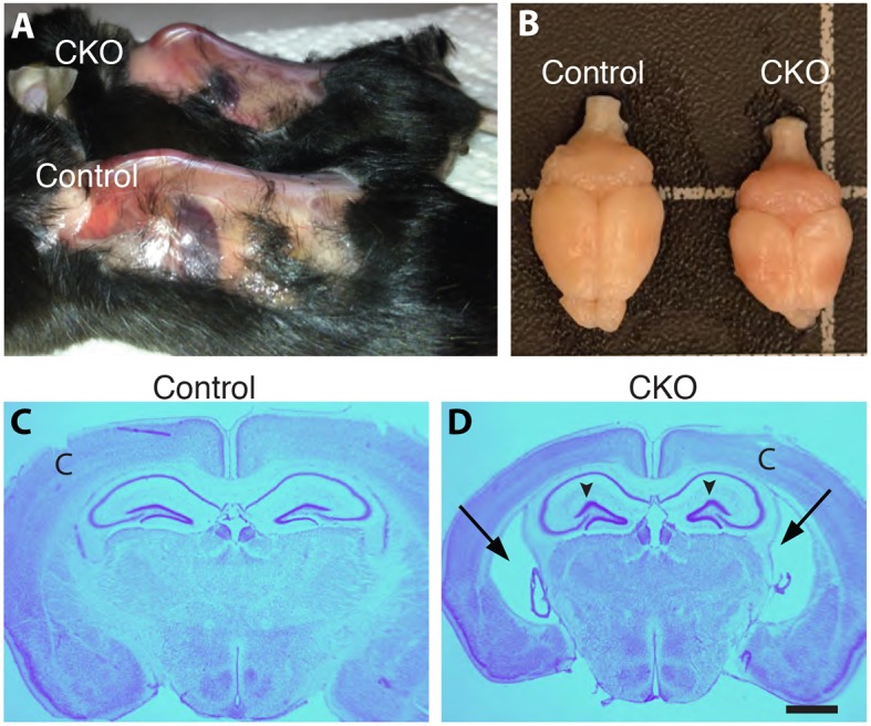
Hereditary sensory and autonomic neuropathies (HSANs) are a genetically and clinically diverse group of disorders defined by peripheral nervous system (PNS) dysfunction. HSAN type III, known as familial dysautonomia (FD), results from a single base mutation in the gene IKBKAP that encodes a scaffolding unit (ELP1) for a multi-subunit complex known as Elongator. Since mutations in other Elongator subunits (ELP2 to ELP4) are associated with central nervous system (CNS) disorders, the goal of this study was to investigate a potential requirement for Ikbkap in the CNS of mice. The sensory and autonomic pathophysiology of FD is fatal, with the majority of patients dying by age 40. While signs and pathology of FD have been noted in the CNS, the clinical and research focus has been on the sensory and autonomic dysfunction, and no genetic model studies have investigated the requirement for Ikbkap in the CNS. Here, we report, using a novel mouse line in which Ikbkap is deleted solely in the nervous system, that not only is Ikbkap widely expressed in the embryonic and adult CNS, but its deletion perturbs both the development of cortical neurons and their survival in adulthood. Primary cilia in embryonic cortical apical progenitors and motile cilia in adult ependymal cells are reduced in number and disorganized. Furthermore, we report that, in the adult CNS, both autonomic and non-autonomic neuronal populations require Ikbkap for survival, including spinal motor and cortical neurons. In addition, the mice developed kyphoscoliosis, an FD hallmark, indicating its neuropathic etiology. Ultimately, these perturbations manifest in a developmental and progressive neurodegenerative condition that includes impairments in learning and memory. Collectively, these data reveal an essential function for Ikbkap that extends beyond the peripheral nervous system to CNS development and function. With the identification of discrete CNS cell types and structures that depend on Ikbkap, novel strategies to thwart the progressive demise of CNS neurons in FD can be developed.

Keywords: Elongator; Familial dysautonomia; Hereditary sensory and autonomic neuropathy.


Conflict of interest statement

Competing interestsThe authors declare no competing or financial interests.

Figures

Fig. 1.
Fig. 1.
Generation of Tuba1a-Cre;Ikbkap CKO mice and expression Tuba1a-cre in the developing CNS. (A) Schematic of conditional mutagenesis in the Ikbkap gene. The β-gal cassette in the knockout Ikbkaptm1a(KOMP)Wtsi allele, which does not express Ikbkap, was removed via Flippase-mediated recombination. In the new IkbkapLoxP allele, Ikbkap is expressed, but exon 4 is flanked by LoxP sites (black triangles) and is excised by Cre in cells that express Tuba in Tuba1a-Cre;IkbkapLoxP/LoxP mice. F and R indicate the locations of primers (forward and reverse, respectively) used to distinguish the CKO Ikbkap allele from the wild-type allele. (B) PCR genotyping. (C,D) Western blots on control and CKO hippocampus, striatum and cortex, and quantification of protein intensity by densitometry demonstrate that IKAP protein is reduced in the striatum and cortex but not in the hippocampus. (E-G) Crossing ROSAmT-mG/mT-mG mice to the Tuba1a-Cre mice reports expression of the Tuba1a-Cre at (E) E12.5 throughout the brain and spinal cord, with detectable expression in the trigeminal nerve, (F) expression in the E17.5 cortex in both the ventricular, subventricular zone and neuronal layers, and expressed in the (G) E17.5 thalamus, but not in the hippocampus. GFP reports Cre expression; Tomato is expressed where Cre is inactive. Arrows in F and G point to the ventricular zone of the cortex; ‘C’, cortex; H, hippocampus; T, thalamus. Scale bars: 1 mm (E); 100 μm (F); 450 μm (G).
Fig. 2.
Fig. 2.
Ikbkap expression in developing and adult CNS. Ikbkap:LacZ reporter mouse shows β-gal expression (A) throughout the neural tube at E8, (B) in the spinal cord motor neuron columns (MN) and ventricular zone (VZ) at E11.5, (C) in both the VZ and subventricular zone (SVZ) and cortical plate in the embryonic cortex at E15.5, with a control shown for comparison in (D), and in (E) several nuclei in the adult medulla (M), including the nucleus tract solitarius (NTS) (*) and cranial motor nuclei (arrows), and (F,G) in the adult cortex (‘C’), hypothalamus (H) and amygdala (‘A’) and hippocampus (* in G). Scale bars: 200 µm (D); 500 µm (E,F).
Fig. 3.
Fig. 3.
Phenotype of Tuba1a-Cre;IC/C mice. (A) CKO mice exhibit kyphosis and (B) microcephaly. (C,D) Nissl staining of coronal sections indicates CKO mice have enlarged lateral ventricles (arrows) and rounded hippocampi with a distorted dentate gyrus lateral blade (arrowheads). Scale bar: 500 µm (D, applies to C). ‘C’, cortex.
Fig. 4.
Fig. 4.
CNS autonomic nuclei neuron number is reduced in the absence of Ikbkap. (A-C) Motor neurons (ChAT positive) in the DMV are reduced in the CKO (see also Table 2). (D-F) nNOS-positive neurons that comprise the intermediolateral cell columns are significantly reduced in number in the CKO thoracic spinal cord. (G-I) PKD2L1-positive cells extend intraluminal buds into the central canal of the spinal cord and are reduced in number and complexity compared to controls. Dashed lines, central canal of spinal cord. Asterisks in A and B denote midline. Error bars denote s.e.m. *P<0.05, unpaired Student's t-test (C,F,I). Scale bar: 30 µm (A,B); 10 µm (D,E,); 20 µm (G,H).
Fig. 5.
Fig. 5.
Neuromuscular junctions in two different muscle groups are denervated in the absence of Ikbkap, and motor neuron numbers are reduced in the CKO spinal cord. Adult (4 month) anterior tibialis muscles (A-E) and erector spinae muscles (F-J) were immunolabeled with α-bungarotoxin (red) to identify cholinergic receptors, and with SV2and 2H3 (green) to label synaptic endings and axons (neurofilaments), respectively. In both muscle types, there was a significant reduction in NMJ innervation, with many terminals being partially innervated (B,H) or completely denervated (D,H). In contrast, at 1 month, neuromuscular junctions are innervated properly in both CKO mice (J) and littermate controls (I). Error bars denote s.e.m. and P-values correspond to an unpaired Student's t-test (*P<0.05). Motor neuron (arrows) numbers (ChAT positive) are reduced in the spinal cords of CKO mice compared to in littermate controls at both 1 month and 4 months. Error bars denote s.e.m. and P-values correspond to an unpaired Student's t-test (*P<0.05). Scale bar: 5 µm (A,B); 2 µm (C,D); 5 µm (G,H); 15 µm (I,J); 20 μm (K,L).
Fig. 6.
Fig. 6.
CNS development requires Ikbkap. (A,B) A 24-h pulse of EdU in E13.5 embryos indicated that progenitor cells produced daughters that migrated in a grossly normal manner to the intermediate zone and cortical plate. (C-E) Immunolabeling at E14.5 for Ki67 and Tuj1 demonstrates the presence of both progenitor cells and neurons, but that the region of the cortex occupied by neurons is thinner in the CKO than in the control brain (*P<0.038; n=3 embryos; Student's t-test). (F-H) In the absence of Ikbkap, the lateral ventricles are expanded in size compared to in control littermates (n=3 embryos; *P<0.002). Scale bar: 50 μm (A-D); 300 μm (F,G).
Fig. 7.
Fig. 7.
Abnormal cilia in both the embryonic and adult lateral ventricles in the absence of Ikbkap. (A-C). Embryonic E14.5 coronal sections were stained with antibodies to adenylate cyclase III (ACIII), and the number of cilia protruding into the lateral ventricles was quantified (n=3 embryos; *P<0.014; Student's t-test). (D-F). In the adult ependymal layer of the lateral ventricle, the number of ependymal cells extending tufts of cilia was significantly reduced in the CKO compared to in the control littermate. Note irregular organization of the ependymal cell layer in the CKO compared to the control. AT, acetylated tubulin. (n=3 brains; *P<0.007; Student's t-test). Scale bar: 15 μm (A,B); 20 μm (D,E).
Fig. 8.
Fig. 8.
CNS behavioral alterations in the absence of Ikbkap. (A) CKO mice show no difference in locomotor activity compared to controls (n=8 per genotype; P>0.05) and (B) no difference in the amount of activity in the center versus at the edges (P>0.05). (C) On the EPM, CKO mice spend a greater proportion of time in the open arms (n=5 control, 6 CKO; *P<0.05) and (D) perform more head dips over the edge of the open arms (n=5 control, 6 CKO; *P<0.05). (E) A similar increase in head-dipping behavior is observed in the cliff avoidance paradigm (n=17 control, 13 CKO; **P<0.005). (F) In the marble burying task, CKO mice bury fewer marbles than controls (n=7 per genotype; *P<0.01). (G) In the Barnes maze, CKO mice fail to use spatial cues to learn the escape location over 7 days of training (n=9 per genotype; *P<0.0001). Error bars denote s.e.m. and P-values correspond to an unpaired Student's t-test.

References

    1. Addis L., Ahn J. W., Dobson R., Dixit A., Ogilvie C. M., Pinto D., Vaags A. K., Coon H., Chaste P., Wilson S. et al. (2015). Microdeletions of ELP4 are associated with language impairment, autism spectrum disorder and mental retardation. Hum. Mutat . 36, 842-850. 10.1002/humu.22816 - DOI - PubMed
    1. Anderson S. L., Coli R., Daly I. W., Kichula E. A., Rork M. J., Volpi S. A., Ekstein J. and Rubin B. Y. (2001). Familial dysautonomia is caused by mutations of the IKAP gene. Am. J. Hum. Genet. 68, 753-758. 10.1086/318808 - DOI - PMC - PubMed
    1. Auer-Grumbach M. (2013). Hereditary sensory and autonomic neuropathies. Handbook Clin. Neurol. 115, 893-906. 10.1016/B978-0-444-52902-2.00050-3 - DOI - PubMed
    1. Axelrod F. B. (2006). A world without pain or tears. Clin. Auton. Res. 16, 90-97. 10.1007/s10286-006-0326-7 - DOI - PubMed
    1. Axelrod F. B., Hilz M. J., Berlin D., Yau P. L., Javier D., Sweat V., Bruehl H. and Convit A. (2010). Neuroimaging supports central pathology in familial dysautonomia. J. Neurol. 257, 198-206. 10.1007/s00415-009-5293-1 - DOI - PubMed

Substances

LinkOut - more resources