. 2017 Feb 21;114(8):1982-1987.
doi: 10.1073/pnas.1617244114. Epub 2017 Feb 6.

Conformational disruption of PI3Kδ regulation by immunodeficiency mutations in PIK3CD and PIK3R1

Affiliations

Conformational disruption of PI3Kδ regulation by immunodeficiency mutations in PIK3CD and PIK3R1

Gillian L Dornan et al. Proc Natl Acad Sci U S A. .

Abstract

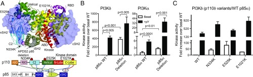
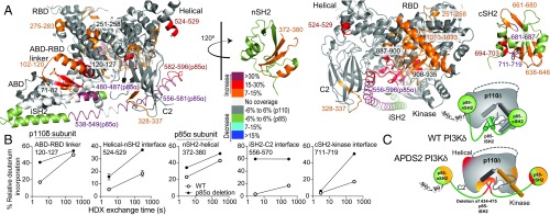
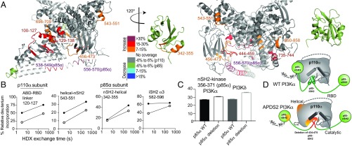
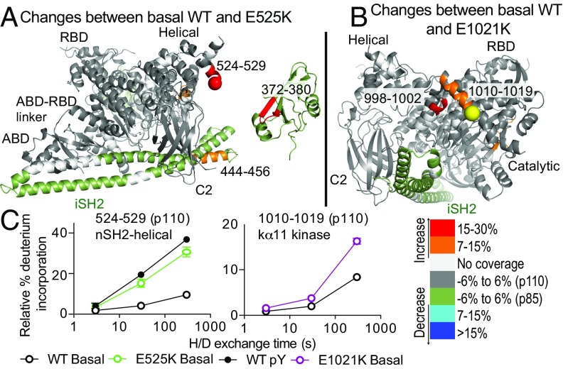
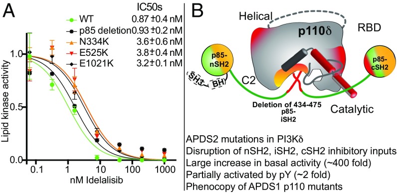
Activated PI3K Delta Syndrome (APDS) is a primary immunodeficiency disease caused by activating mutations in either the leukocyte-restricted p110δ catalytic (PIK3CD) subunit or the ubiquitously expressed p85α regulatory (PIK3R1) subunit of class IA phosphoinositide 3-kinases (PI3Ks). There are two classes of APDS: APDS1 that arises from p110δ mutations that are analogous to oncogenic mutations found in the broadly expressed p110α subunit and APDS2 that occurs from a splice mutation resulting in p85α with a central deletion (Δ434-475). As p85 regulatory subunits associate with and inhibit all class IA catalytic subunits, APDS2 mutations are expected to similarly activate p110α, β, and δ, yet APDS2 largely phenocopies APDS1 without dramatic effects outside the immune system. We have examined the molecular mechanism of activation of both classes of APDS mutations using a combination of biochemical assays and hydrogen-deuterium exchange mass spectrometry. Intriguingly, we find that an APDS2 mutation in p85α leads to substantial basal activation of p110δ (>300-fold) and disrupts inhibitory interactions from the nSH2, iSH2, and cSH2 domains of p85, whereas p110α is only minimally basally activated (∼2-fold) when associated with mutated p85α. APDS1 mutations in p110δ (N334K, E525K, E1021K) mimic the activation mechanisms previously discovered for oncogenic mutations in p110α. All APDS mutations were potently inhibited by the Food and Drug Administration-approved p110δ inhibitor idelalisib. Our results define the molecular basis of how PIK3CD and PIK3R1 mutations result in APDS and reveal a potential path to treatment for all APDS patients.

Keywords: HDX-MS; PI3K/AKT; PIK3CD; PIK3R1; phosphoinositides.


Conflict of interest statement

Conflict of interest statement: C.L.L. collaborates with Novartis on related studies.

Figures

Fig. 1.
Fig. 1.
APDS1 and APDS2 mutants lead to increased basal and pY-activated lipid kinase activity compared with WT. (A) The location of APDS mutations in p110δ/p85α is mapped on a model of the PI3Kδ complex based on the structure of the p110δ/p85α iSH2 complex [Protein Data Bank (PDB): 5DXU (37)] with the nSH2 modeled from the PI3Kα structure [PDB: 3HHM (38)] and the cSH2 modeled from the PI3Kβ structure [PDB: 2Y3A (13)]. The nSH2 and cSH2 are shown as transparent, with APDS1 mutations shown as pink spheres. The APDS2 truncation in the iSH2 is white. Full molecular details of the APDS mutants are shown in SI Appendix, Fig. S1. A domain schematic of p110δ/p85α is included with inhibitory interfaces highlighted. (B) Fold activation of APDS2 mutation in the context of PI3Kα and PI3Kδ. Lipid kinase assays of WT PI3Kδ or PI3Kα with WT p85α or the APDS2 p85α splice variant [p85α (Δ434–475)] in the presence (+pY) or absence (basal) of a stimulating RTK-derived phosphopeptide (1 μM). Specific activity was normalized to WT (p110δ/α-p85α). (C) Fold activation of APDS1 mutations in p110δ in the presence (+pY) or absence (basal) of a stimulating RTK-derived phosphopeptide. Assays measured the production of ADP in the presence of 0.1–100 nM of enzyme, 100 μM ATP, and 5% PIP2/95% PS vesicles. Kinase assays were performed in triplicate (error shown as SD; n = 3).
Fig. 2.
Fig. 2.
HDX-MS reveals that APDS2 mutation in p85α leads to disruption of inhibitory interactions in PI3Kδ. (A) Peptides in p110δ and p85α that showed differences in HDX both greater than 0.7 Da and 7% in the APDS2 p85α mutation compared with the WT are highlighted on the structural model from Fig. 1A according to the legend. nSH2 and cSH2 are shown disconnected from the catalytic subunit as the HDX data suggest that these interfaces are disrupted. (B) Time course of deuterium incorporation for a selection of peptides in both p110δ and p85α with differences in HDX in the APDS2 p85α mutant (error shown as SD; n = 3). (C) Schematic model of WT PI3Kδ and conformational changes that occur in the complex with the APDS2 splice variant of p85.
Fig. 3.
Fig. 3.
HDX-MS reveals that APDS2 mutation in p85α leads to partial disruption of inhibitory interactions in PI3Kα. (A) Peptides in p110α and p85α that showed differences in HDX both greater than 0.7 Da and 7% in the APDS2 p85α mutation compared with the WT are highlighted on the structure of p110α bound to the nSH2 and iSH2 of p85α [PDB: 3HHM (38)]. The nSH2 domain is shown disconnected from the catalytic subunit, as the HDX data suggest that this interface is partially disrupted. (B) Time course of deuterium incorporation for a selection of peptides in both p110α and p85α with HDX differences in the APDS2 PIK3R1 mutant. (C) HDX exchange levels for the APDS2 complex of PI3Kα and PI3Kδ compared with WT for a peptide in the nSH2 of p85α located at the nSH2–kinase interface at the 300-s time point. Error bars in all graphs represent SD (n = 3). (D) Schematic model of WT PI3Kα and conformational changes that occur in the complex with the APDS2 splice variant of p85α.
Fig. 4.
Fig. 4.
HDX-MS reveals that APDS1 mutations in p110δ mimic the activated form of WT PI3Kδ. (A and B) Peptides in p110δ and p85α with HDX differences both greater than 0.7 Da and 7% in the APDS1 mutants E525K and E1021K in PIK3CD compared with the WT are highlighted on the structural model as in Fig. 2. (C) Time course of deuterium incorporation for a selection of peptides in both p110δ and p85α under a variety of conditions (basal, pY-phosphopeptide bound) with HDX differences in the APDS1 mutants. Error bars represent SD (n = 3).
Fig. 5.
Fig. 5.
Inhibition of APDS1 and APDS2 mutants by the potent PI3Kδ inhibitor idelalisib and model for activation of PI3Kδ and PI3Kα by APDS2 mutations in PIK3R1. (A) Inhibition of WT, APDS1, and APDS2 complexes of PI3Kδ by the potent PI3Kδ inhibitor idelalisib. Lipid kinase activity was normalized to kinase activity in the absence of inhibitors. IC50 values were generated from triplicate independent inhibitor dilutions, and error is shown as SD. (B) Summary of the proposed mechanism for activation of the APDS2 PI3Kδ complex mapped on a schematic model with conformational changes and kinase activity data summarized.

References

    1. Burke JE, Williams RL. Synergy in activating class I PI3Ks. Trends Biochem Sci. 2015;40(2):88–100. - PubMed
    1. Vanhaesebroeck B, Guillermet-Guibert J, Graupera M, Bilanges B. The emerging mechanisms of isoform-specific PI3K signalling. Nat Rev Mol Cell Biol. 2010;11(5):329–341. - PubMed
    1. Samuels Y, et al. High frequency of mutations of the PIK3CA gene in human cancers. Science. 2004;304(5670):554. - PubMed
    1. Dyment DA, et al. FORGE Canada Consortium Mutations in PIK3R1 cause SHORT syndrome. Am J Hum Genet. 2013;93(1):158–166. - PMC - PubMed
    1. Lucas CL, Chandra A, Nejentsev S, Condliffe AM, Okkenhaug K. PI3Kδ and primary immunodeficiencies. Nat Rev Immunol. 2016;16(11):702–714. - PMC - PubMed

Publication types

MeSH terms

Supplementary concepts