

Crystal structure of the adenosine A2A receptor bound to an antagonist reveals a potential allosteric pocket
- PMID: 28167788
- PMCID: PMC5338372
- DOI: 10.1073/pnas.1621423114
Crystal structure of the adenosine A2A receptor bound to an antagonist reveals a potential allosteric pocket
Abstract
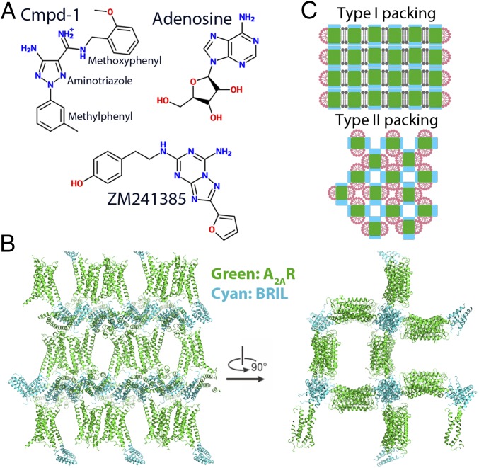
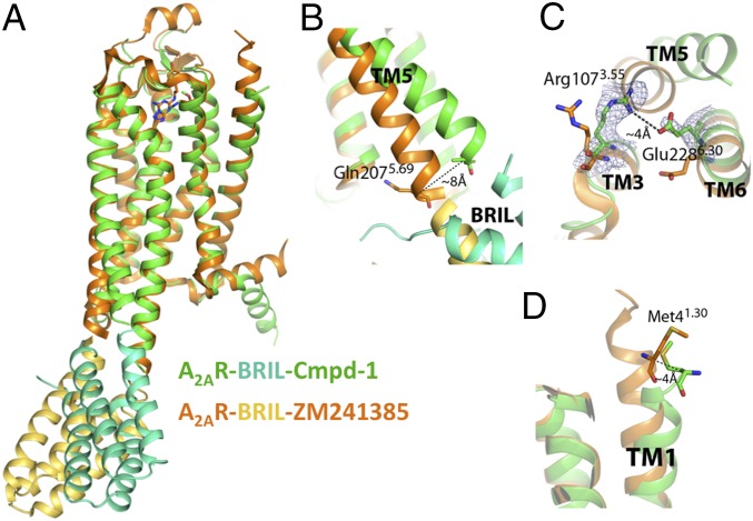
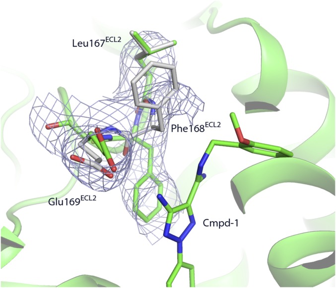
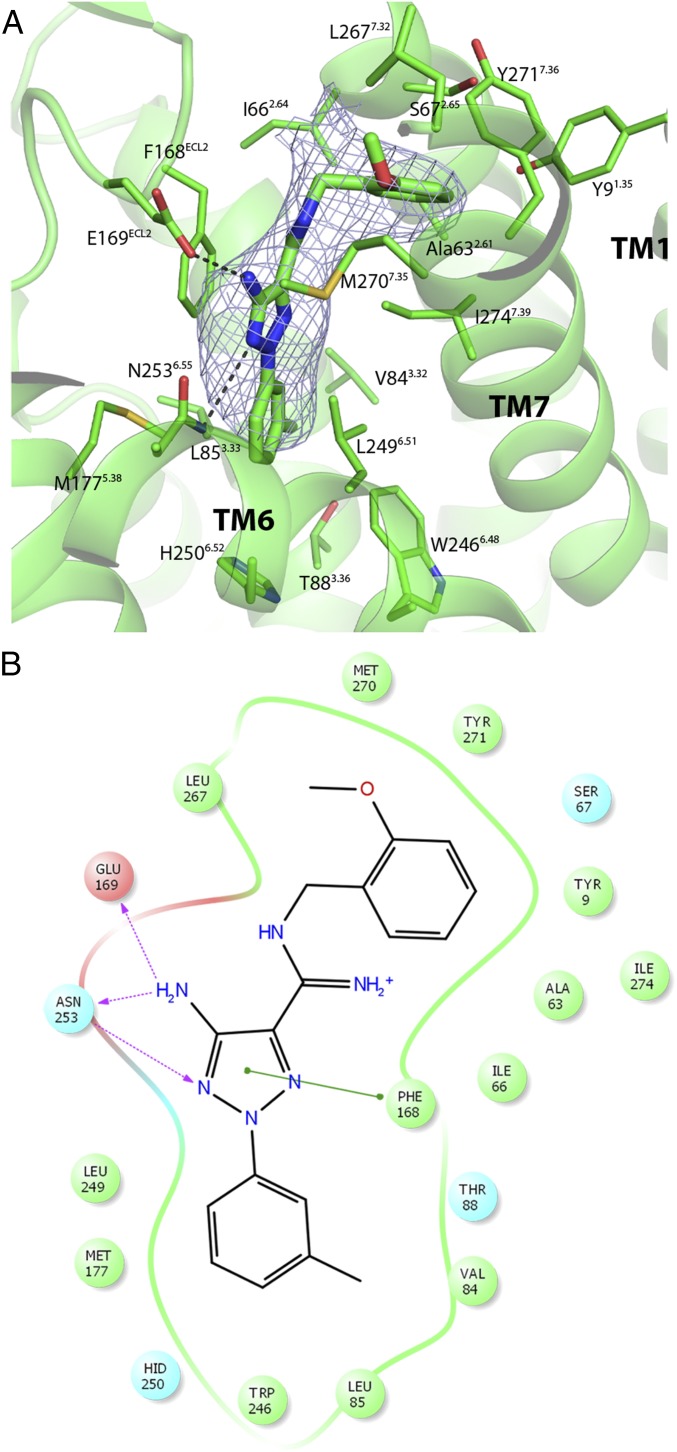
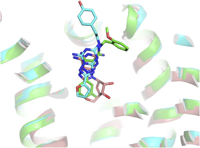
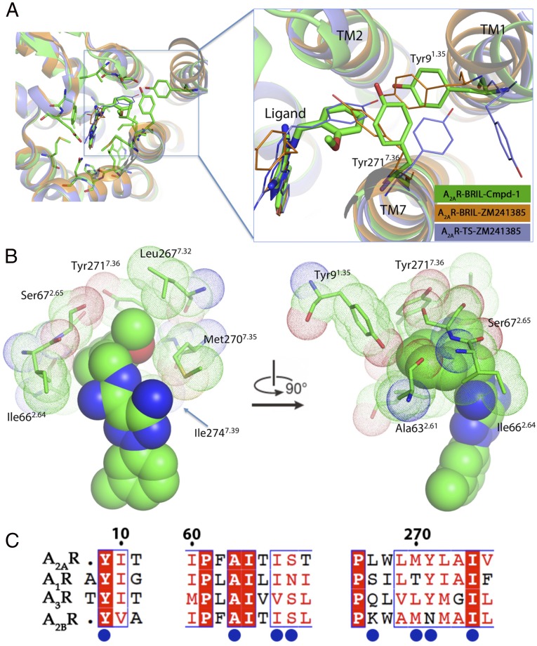
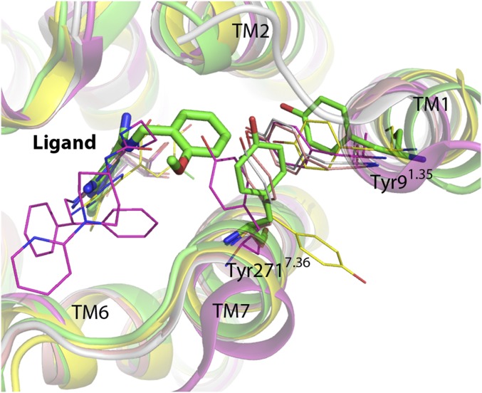
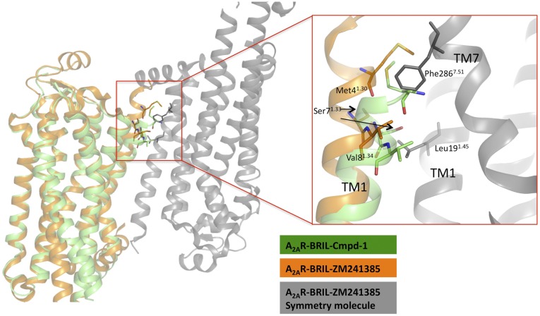
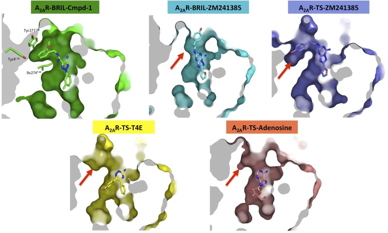
The adenosine A2A receptor (A2AR) has long been implicated in cardiovascular disorders. As more selective A2AR ligands are being identified, its roles in other disorders, such as Parkinson's disease, are starting to emerge, and A2AR antagonists are important drug candidates for nondopaminergic anti-Parkinson treatment. Here we report the crystal structure of A2A receptor bound to compound 1 (Cmpd-1), a novel A2AR/N-methyl d-aspartate receptor subtype 2B (NR2B) dual antagonist and potential anti-Parkinson candidate compound, at 3.5 Å resolution. The A2A receptor with a cytochrome b562-RIL (BRIL) fusion (A2AR-BRIL) in the intracellular loop 3 (ICL3) was crystallized in detergent micelles using vapor-phase diffusion. Whereas A2AR-BRIL bound to the antagonist ZM241385 has previously been crystallized in lipidic cubic phase (LCP), structural differences in the Cmpd-1-bound A2AR-BRIL prevented formation of the lattice observed with the ZM241385-bound receptor. The crystals grew with a type II crystal lattice in contrast to the typical type I packing seen from membrane protein structures crystallized in LCP. Cmpd-1 binds in a position that overlaps with the native ligand adenosine, but its methoxyphenyl group extends to an exosite not previously observed in other A2AR structures. Structural analysis revealed that Cmpd-1 binding results in the unique conformations of two tyrosine residues, Tyr91.35 and Tyr2717.36, which are critical for the formation of the exosite. The structure reveals insights into antagonist binding that are not observed in other A2AR structures, highlighting flexibility in the binding pocket that may facilitate the development of A2AR-selective compounds for the treatment of Parkinson's disease.
Keywords: A2A adenosine receptor; GPCR; Parkinson’s disease; allosteric; structure.
Conflict of interest statement
Conflict of interest statement: The research was performed by ConfometRx, Inc., cofounded by T.S.K. and B.K.K., in collaboration with UCB Pharma.
Figures
Similar articles
-
Design and development of 1,3,5-triazine-thiadiazole hybrids as potent adenosine A2A receptor (A2AR) antagonist for benefit in Parkinson's disease.Neurosci Lett. 2020 Sep 14;735:135222. doi: 10.1016/j.neulet.2020.135222. Epub 2020 Jun 30. Neurosci Lett. 2020. PMID: 32619652
-
Novel approaches for targeting the adenosine A2A receptor.Expert Opin Drug Discov. 2015 Jan;10(1):63-80. doi: 10.1517/17460441.2015.971006. Epub 2014 Oct 14. Expert Opin Drug Discov. 2015. PMID: 25311639 Review.
-
Discovery of potent adenosine A2a antagonists as potential anti-Parkinson disease agents. Non-linear QSAR analyses integrated with pharmacophore modeling.Chem Biol Interact. 2016 Jul 25;254:93-101. doi: 10.1016/j.cbi.2016.05.023. Epub 2016 May 20. Chem Biol Interact. 2016. PMID: 27216633
-
Molecular Basis of Ligand Dissociation from the Adenosine A2A Receptor.Mol Pharmacol. 2016 May;89(5):485-91. doi: 10.1124/mol.115.102657. Epub 2016 Feb 12. Mol Pharmacol. 2016. PMID: 26873858
-
Allosteric Interactions between Adenosine A2A and Dopamine D2 Receptors in Heteromeric Complexes: Biochemical and Pharmacological Characteristics, and Opportunities for PET Imaging.Int J Mol Sci. 2021 Feb 9;22(4):1719. doi: 10.3390/ijms22041719. Int J Mol Sci. 2021. PMID: 33572077 Free PMC article. Review.
Cited by
-
Computational Methods for the Discovery and Optimization of TAAR1 and TAAR5 Ligands.Int J Mol Sci. 2024 Jul 27;25(15):8226. doi: 10.3390/ijms25158226. Int J Mol Sci. 2024. PMID: 39125796 Free PMC article. Review.
-
Metal Chelation Therapy and Parkinson's Disease: A Critical Review on the Thermodynamics of Complex Formation between Relevant Metal Ions and Promising or Established Drugs.Biomolecules. 2019 Jul 9;9(7):269. doi: 10.3390/biom9070269. Biomolecules. 2019. PMID: 31324037 Free PMC article. Review.
-
Could confounding the allosteric communication of biotic machinery be an alternative path to antibiotics?Proc Natl Acad Sci U S A. 2020 Apr 14;117(15):8222-8224. doi: 10.1073/pnas.2002666117. Epub 2020 Mar 20. Proc Natl Acad Sci U S A. 2020. PMID: 32198204 Free PMC article. No abstract available.
-
3D-e-Chem: Structural Cheminformatics Workflows for Computer-Aided Drug Discovery.ChemMedChem. 2018 Mar 20;13(6):614-626. doi: 10.1002/cmdc.201700754. Epub 2018 Feb 14. ChemMedChem. 2018. PMID: 29337438 Free PMC article.
-
Neutrophil Signaling That Challenges Dogmata of G Protein-Coupled Receptor Regulated Functions.ACS Pharmacol Transl Sci. 2020 Mar 11;3(2):203-220. doi: 10.1021/acsptsci.0c00004. eCollection 2020 Apr 10. ACS Pharmacol Transl Sci. 2020. PMID: 32296763 Free PMC article. Review.
References
-
- de Lera Ruiz M, Lim YH, Zheng J. Adenosine A2A receptor as a drug discovery target. J Med Chem. 2014;57(9):3623–3650. - PubMed
-
- Pinna A, Wardas J, Simola N, Morelli M. New therapies for the treatment of Parkinson’s disease: Adenosine A2A receptor antagonists. Life Sci. 2005;77(26):3259–3267. - PubMed
-
- Bara-Jimenez W, et al. Adenosine A(2A) receptor antagonist treatment of Parkinson’s disease. Neurology. 2003;61(3):293–296. - PubMed
MeSH terms
Substances
LinkOut - more resources
Full Text Sources
Other Literature Sources
Medical
Research Materials
