. 2018 Mar;23(3):747-758.
doi: 10.1038/mp.2016.253. Epub 2017 Feb 7.

Autism-associated Dyrk1a truncation mutants impair neuronal dendritic and spine growth and interfere with postnatal cortical development

Affiliations

Autism-associated Dyrk1a truncation mutants impair neuronal dendritic and spine growth and interfere with postnatal cortical development

T Dang et al. Mol Psychiatry. 2018 Mar.

Abstract

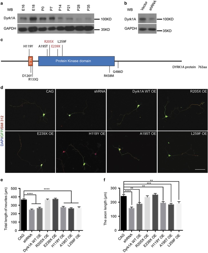
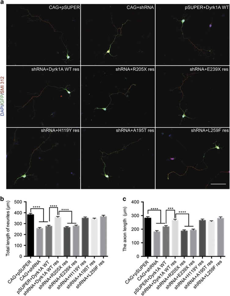
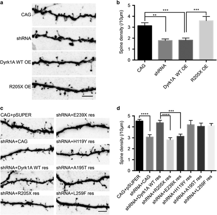
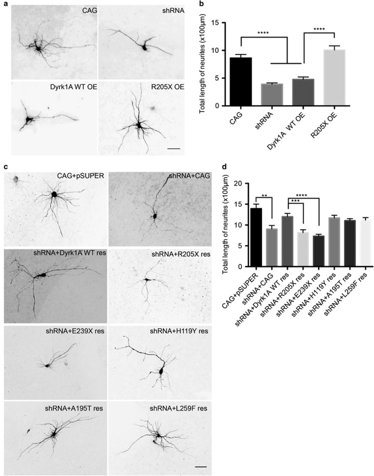
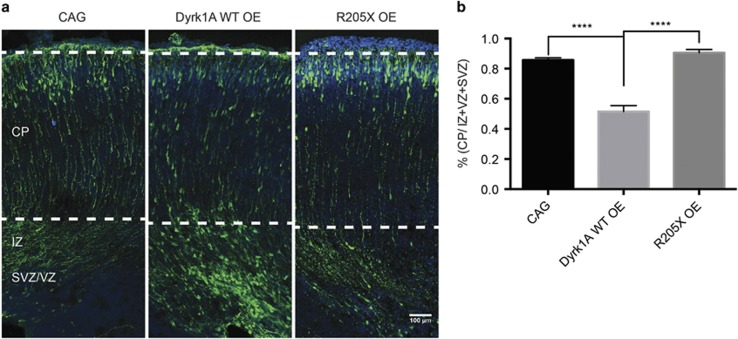
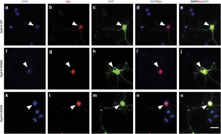
Autism is a prevailing neurodevelopmental disorder with a large genetic/genomic component. Recently, the dual-specificity tyrosine-(Y)-phosphorylation-regulated kinase 1 A (DYRK1A) gene was implicated as a risk factor for autism spectrum disorder (ASD). We identified five DYRK1A variants in ASD patients and found that the dose of DYRK1A protein has a crucial role in various aspects of postnatal neural development. Dyrk1a loss of function and gain of function led to defects in dendritic growth, dendritic spine development and radial migration during cortical development. Importantly, two autism-associated truncations, R205X and E239X, were shown to be Dyrk1a loss-of-function mutants. Studies of the truncated Dyrk1a mutants may provide new insights into the role of Dyrk1a in brain development, as well as the role of Dyrk1a loss of function in the pathophysiology of autism.


Conflict of interest statement

The authors declare no conflict of interest.

Figures

Figure 1
Figure 1
Wild-type (WT) Dyrk1a (dual-specificity tyrosine-(Y)-phosphorylation-regulated kinase 1 A) overexpression reduced total neurite and axon length of neurons in primary cultured mouse cortical neurons. (a) The Dyrk1a expression pattern in the cerebral cortex during the embryonic and postnatal period. (b) Specific knockdown of endogenous Dyrk1a in the mouse cortical neurons. Immunoblotting of the knockdown using specific short hairpin RNA (shRNA) against endogenous DYRK1A protein. The protein marker ladders are shown on the right. (c) Representation of the specific locations of missense variants related to autism in the DYRK1A protein structure, with the two truncations highlighted in red. The five variants located above the protein structure were included in the additional functional study. The smaller orange bar shows the NLS (bipartite nuclear localization signal) motif, and the larger bar (blue) represents the protein kinase domain. The black thread shows the other amino acids making up DYRK1A. (d) The specific cellular morphology of neurons expressing the CAG vector (as a control), Dyrk1a RNAi, Dyrk1a WT overexpression, the two truncations (R205X and E239X) and the three missense mutations (H119Y, A195T and L259F). All neurons were co-labeled with 4,6-diamidino-2-phenylindole (DAPI; to identify nuclei), green fluorescent protein (GFP; to identify overall neuronal morphology) and SMI 312 (an axonal marker). Scale bar 50 μm. (e, f) The statistical results for the total neurites (e) and axon length (f) between specific cell types. The bar graph shows the mean value±s.e.m.; approximately 70 cells from three independent experiments were counted during the statistical analysis. Unpaired t-test statistical method was used. **P<0.01, ***P<0.001, ****P<0.0001.
Figure 2
Figure 2
Wild-type (WT) Dyrk1a (dual-specificity tyrosine-(Y)-phosphorylation-regulated kinase 1 A) rescues the phenotype of decreased neurites and axon length in cultured mouse cortical neurons following Dyrk1a short hairpin RNA (shRNA) knockdown. (a) The specific morphology of neurons expressing the vectors (CAG and pSUPER), shRNA, WT Dyrk1a and WT or mutant forms of Dyrk1a along with the shRNA. All neurons were co-labeled with 4,6-diamidino-2-phenylindole (DAPI), green fluorescent protein (GFP) and SMI 312 (to distinguish axons from total neurites). Scale bar 50 μm. (b, c) The statistical results for the total neurites (b) and axon length (c) between specific cell types. The bar graph shows the mean value±s.e.m.; approximately 60 cells from three independent experiments were counted, and unpaired t-tests were used for the statistical analysis. ***P<0.001, ****P<0.0001.
Figure 3
Figure 3
Dyrk1a (dual-specificity tyrosine-(Y)-phosphorylation-regulated kinase 1 A) dosage imbalance reduced dendritic spine density in rat hippocampal slice culture, and wild-type (WT) Dyrk1a rescued the phenotype caused by Dyrk1a knockdown. (a, c) Morphology of the spine density under specific conditions with the expression of different plasmids, including CAG vector, Dyrk1a short hairpin RNA (shRNA), Dyrk1a WT, R205X (a) and shRNA rescue using WT or mutant Dyrk1a (c). Scale bar 5 μm. (b, d) Statistical analysis of the spine density (per 10 μm) in specific types of neurons. Forty-five neurons from three independent experiments were counted, and the individual neuronal spine density was determined. Unpaired t-tests were performed for the statistical analysis to compare each condition. **P<0.01, ***P<0.001, ****P<0.0001.
Figure 4
Figure 4
Dyrk1a (dual-specificity tyrosine-(Y)-phosphorylation-regulated kinase 1 A) inhibited total neurite outgrowth in rat hippocampal neurons, and short hairpin RNA (shRNA)-mediated loss of function was rescued by wild-type Dyrk1a. (a, c) The morphology of neurons in each condition expressing the indicated plasmids. (a) Neurons expressed CAG vector, Dyrk1a short hairpin RNA (shRNA), Dyrk1a wild-type (WT) and R205X. (c) Neurons expressed specific plasmids for the rescue conditions, including WT and the five mutant forms of Dyrk1a plus Dyrk1a shRNA. (b, d) Statistical analysis of total neurite length in overexpression (b) and rescue conditions (d). Scale bar 50 μm. Approximately 50 neurons from three independent experiments were counted, and the total neurite length of each cell was determined. Unpaired t-tests were performed for the statistical analysis. **P<0.01, ***P<0.001, ****P<0.0001.
Figure 5
Figure 5
Dyrk1a (dual-specificity tyrosine-(Y)-phosphorylation-regulated kinase 1 A) wild-type (WT) overexpression delayed neuronal migration in mouse embryonic cortical development. (a) Cortical migration in each condition, including CAG (left), Dyrk1a WT overexpression (middle) and R205X (right). (b) Quantification of panel (a). The green fluorescent protein (GFP)-positive cells in the cortical plate (CP), intermediate zone (IZ), subventricular zone (SVZ) and ventricular zone (VZ) were counted, and the ratio of GFP-positive cells in the CP/(IZ and VZ/SVZ) was analyzed statistically using unpaired t-tests. P0 mice were collected in three independent experiments, including CAG (n=7), Dyrk1A WT (n=6) and R205X (n=5) mice. ****P<0.0001. Scale bar 100 μm.
Figure 6
Figure 6
The two Dyrk1a (dual-specificity tyrosine-(Y)-phosphorylation-regulated kinase 1 A) truncations did not disturb Dyrk1a subcellular localization. (ae) Neurons expressing Dyrk1a wild-type (WT) were labeled with 4,6-diamidino-2-phenylindole (DAPI; to identify nuclei), a Myc epitope tag (to identify the precise location of the expressed protein) and green fluorescent protein (GFP; to identify overall neuronal morphology). Merge 1 shows DAPI and Myc co-labeling, and merge 2 shows DAPI, Myc and GFP labeling. Panels (fj) and (ko) depict the two nonsense mutants, respectively. Both R205X and E239X, as well as Dyrk1a WT, localized to the nucleus. The images were merged using the ImageJ software. Scale bar 20 μm.

References

    1. Blenner S, Reddy A, Augustyn M. Diagnosis and management of autism in childhood. BMJ 2011; 343: d6238. - PubMed
    1. Zafeiriou DI, Ververi A, Dafoulis V, Kalyva E, Vargiami E. Autism spectrum disorders: the quest for genetic syndromes. Am J Med Genet B Neuropsychiatr Genet 2013; 162B: 327–366. - PubMed
    1. Geschwind DH, Levitt P. Autism spectrum disorders: developmental disconnection syndromes. Curr Opin Neurobiol 2007; 17: 103–111. - PubMed
    1. Geschwind DH. Autism: many genes, common pathways? Cell 2008; 135: 391–395. - PMC - PubMed
    1. Betancur C. Etiological heterogeneity in autism spectrum disorders: more than 100 genetic and genomic disorders and still counting. Brain Res 2011; 1380: 42–77. - PubMed

Publication types

MeSH terms

Substances