. 2017 Jan 23:10:8.
doi: 10.3389/fnmol.2017.00008. eCollection 2017.

Parvalbumin-Neurons of the Ventrolateral Hypothalamic Parvafox Nucleus Receive a Glycinergic Input: A Gene-Microarray Study

Affiliations

Parvalbumin-Neurons of the Ventrolateral Hypothalamic Parvafox Nucleus Receive a Glycinergic Input: A Gene-Microarray Study

Viktoria Szabolcsi et al. Front Mol Neurosci. .

Abstract

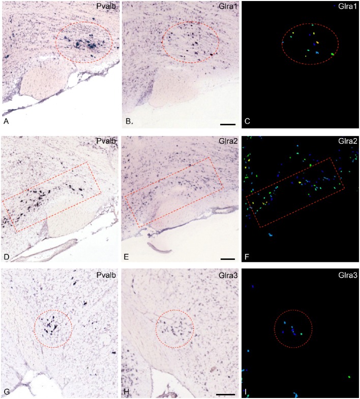
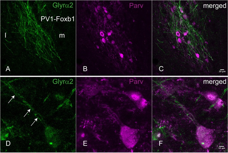
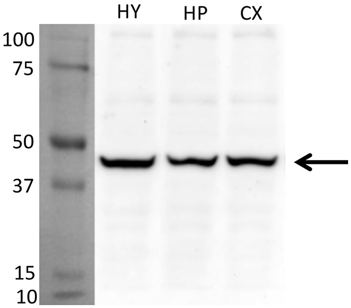
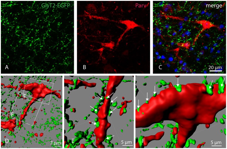
The ventrolateral hypothalamic parvafox (formerly called PV1-Foxb1) nucleus is an anatomical entity of recent discovery and unknown function. With a view to gaining an insight into its putative functional role(s), we conducted a gene-microarray analysis and, armed with the forthcoming data, controlled the results with the Allen databases and the murine BrainStars (B*) database. The parvafox nucleus was specifically sampled by laser-capture microdissection and the transcriptome was subjected to a microarray analysis on Affymetrix chips. Eighty-two relevant genes were found to be potentially more expressed in this brain region than in either the cerebral cortex or the hippocampus. When the expression patterns of these genes were counterchecked in the Allen-Database of in-situ hybridizations and in the B*-microarray database, their localization in the parvafox region was confirmed for thirteen. For nine novel genes, which are particularly interesting because of their possible involvement in neuromodulation, the expression was verified by quantitative real time-PCR. Of particular functional importance may be the occurrence of glycine receptors, the presence of which indicates that the activity of the parvafox nucleus is under ascending inhibitory control.

Keywords: Allen Brain Atlas Database of in-situ hybridizations (ABA-ISH); BrainStars (B*) microarrays; GlyT2-EGFP; PV1-Foxb1; glycine receptor α2; laser capture microdissection; parvafox; parvalbumin.


Figures

Figure 1
Figure 1
Expression of the mRNA for the glycine-receptor subunits α-1, α-2 and α-3 in the parvafox nucleus as found in the ABA-ISH. (A,D,G) In situ hybridization-images for Pvalb, downloaded from the ABA website, defining the location of the parvafox nucleus (surrounded by a red circle or rectangle). (B,E,H): The mRNAs for the glycine-receptor subunits α-1, α-2, and α-3 are expressed within the confines of the parvafox nucleus. In (C,F,I) the expression level of Glra1, Glra2, and Glra3 is visualized with pseudocolors. (A–F) are sagittal sections and (G–I) coronal sections. (Image credit: Allen Institute.) Scale bars represent 100 μm.
Figure 2
Figure 2
Immunohistochemical localization of the Glyrα2 receptor in the parvafox nucleus. In overview pictures, the Glyrα2 immunoreactive sites in the hypothalamus are concentrated in the ventrolateral hypothalamus and the mammillary nuclei. At low magnification, a meshwork of Glyrα2 immunoreactive fibers (A) is intermingled with Parv-immunoreactive neurons (B,C). At higher magnification, Glyrα2 immunoreactivity decorates the dendrites (arrows, D) and cell body of Parv-positive neurons (E,F).
Figure 3
Figure 3
Detection of Glyrα2 on hypothalamic, hippocampal and cortical extracts by immunoblot. The antiserum against Glyrα2 detects a single band below 50 KDa (arrow) in an immunoblot of hypothalamic, hippocampal and cerebral cortical extracts submitted to SDS-gel electrophoresis. (HY, hypothalamus; HP, hippocampus; CX, cortex).
Figure 4
Figure 4
Localization of GlyT2-EGFP-positive axons and axon terminals in the parvafox nucleus. Confocal laser micrographs of double-immunohistochemistry for GFP (green, A) and Parv (red, B) performed on brain sections of transgenic GlyT2-EGFP mice revealed the presence of several axons and axon terminals in the hypothalamic region encompassing the parvafox nucleus. The merged image (C) shows GlyT2-EGFP-positive terminals around the perikaryon and the dendrites of a Parv-positive neuron. Three-dimensional reconstruction by Imaris (D) reveals several EGFP-positive terminals on the dendrite (E, see arrows), as well as on the cell body (F, see arrows) of the Parv-neuron.

References

    1. Akaike N., Kaneda M. (1989). Glycine-gated chloride current in acutely isolated rat hypothalamic neurons. J. Neurophysiol. 62, 1400–1409. - PubMed
    1. Barreiro-Iglesias A., Mysiak K. S., Adrio F., Rodicio M. C., Becker C. G., Becker T., et al. . (2013). Distribution of glycinergic neurons in the brain of glycine transporter-2 transgenic Tg(glyt2:Gfp) adult zebrafish: relationship to brain-spinal descending systems. J. Comp. Neurol. 521, 389–425. 10.1002/cne.23179 - DOI - PubMed
    1. Bilella A., Alvarez-Bolado G., Celio M. R. (2014). Coaxiality of Foxb1- and parvalbumin-expressing neurons in the lateral hypothalamic PV1-nucleus. Neurosci. Lett. 566, 111–114. 10.1016/j.neulet.2014.02.028 - DOI - PubMed
    1. Bilella A., Alvarez-Bolado G., Celio M. R. (2016). The Foxb1-expressing neurons of the ventrolateral hypothalamic parvafox nucleus project to defensive circuits. J. Comp. Neurol. 524, 2955–2981. 10.1002/cne.24057 - DOI - PubMed
    1. Brackmann M., Zhao C., Schmieden V., Braunewell K. H. (2004). Cellular and subcellular localization of the inhibitory glycine receptor in hippocampal neurons. Biochem. Biophys. Res. Commun. 324, 1137–1142. 10.1016/j.bbrc.2004.09.172 - DOI - PubMed