. 2017 Jan 24:8:35.
doi: 10.3389/fimmu.2017.00035. eCollection 2017.

Mouse Liver Sinusoidal Endothelium Eliminates HIV-Like Particles from Blood at a Rate of 100 Million per Minute by a Second-Order Kinetic Process

Affiliations

Mouse Liver Sinusoidal Endothelium Eliminates HIV-Like Particles from Blood at a Rate of 100 Million per Minute by a Second-Order Kinetic Process

Jessica M Mates et al. Front Immunol. .

Abstract

We crafted human immunodeficiency virus (HIV)-like particles of diameter about 140 nm, which expressed two major HIV-1 proteins, namely, env and gag gene products, and used this reagent to simulate the rate of decay of HIV from the blood stream of BALB/c male mice. We found that most (~90%) of the particles were eliminated (cleared) from the blood by the liver sinusoidal endothelial cells (LSECs), the remainder from Kupffer cells; suggesting that LSECs are the major liver scavengers for HIV clearance from blood. Decay was rapid with kinetics suggesting second order with respect to particles, which infers dimerization of a putative receptor on LSEC. The number of HIV-like particles required for saturating the clearance mechanism was approximated. The capacity for elimination of blood-borne HIV-like particles by the sinusoid was 112 million particles per minute. Assuming that the sinusoid endothelial cells were about the size of glass-adherent macrophages, then elimination capacity was more than 540 particles per hour per endothelial cell.

Keywords: Kupffer cell; clearance; endocytosis; liver sinusoidal endothelial cell; pinocytosis.


Figures

Figure 1
Figure 1
HIV-like particles are cleared rapidly from murine circulation. Approximately 2 × 1010 HIV-like particles were intravenously infused by tail vein. The concentration of particles remaining in blood of the suborbital plexus over time was estimated using a p24 ELISA. (A) Shows a plot of mean ± SD of the decay curves. The curve was drawn using asymmetric sigmoidal five parameters simulation to smooth the connection of data points. (B) Shows decay curves of three different mice illustrating the splay of the SD in (A); one high, one mid-level, and one low. (C) Shows a log–linear plot of the data illustrating no straight line. (D) Plots the data in log–log fashion to reveal a straight line of pseudo second order kinetics. (E) Shows a reciprocal plot of the same data, showing also a straight line. The 30-min data points that did not fall on the straight line are not shown; they represent less than 3% of the dose. Each data point represents mean ± SD of 26 BALB/c wild-type mice.
Figure 2
Figure 2
The liver is the major organ clearing Cy3-HIV-like particles from blood. Mice were infused with 1010 Cy3-human immunodeficiency virus-like particles and, after 10 min of infusion, the Cy3 fluorescence was quantified in various organs as described in M&M. The bar graph expresses the percentage means and SDs of total Cy3 fluorescence that was recovered from the six organs studied. The asterisk represents data points where the p-Values were determined to be less than 0.05 using Student’s t-test.
Figure 3
Figure 3
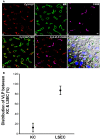
The majority of human immunodeficiency virus (HIV)-like particles cleared by liver is localized to the liver sinusoidal endothelial cell (LSEC). (A) Four-color fluorescence microscopic images of 5 µm liver sections, 3 min after intravenous infusion of 2 × 1010 Cy3-HIV-like particles. (a) Red puncta show Cy3-HIV-like particles. (b) rabbit anti-mannose receptor (CD206) labeling of LSEC shown in green. (c) rat mab F4/80 labeling of KC shown in magenta. (d) Cy3-VLP (red) merged with LSEC marker. (e) Cy3-HIV-like particles (red) merged with KC marker. (f) Merged image showing Cy3-HIV-like particles (red), LSEC marker (green), and KC marker (magenta) plus DIC and DAPI staining of the nuclei (blue). Panels shown are representative of 160 images from three different mice. The scale bar in panel (c) signifies 5 µm. (B) Quantified association of HIV-like particles with cells of the liver. All HIV-like particles in the liver were associated with either LSEC or KC, indicating no association with hepatocytes. The total HIV-like particles was calculated as the pixel area × mean fluorescence intensity (red). HIV-like particles associated with the KC was subtracted from the total HIV-like particle to calculate LSEC association of HIV-like particle. The graph represents the mean ± SD of KC- and LSEC-association within liver. One hundred sixty images, roughly 50 from each of the three mice, were examined. The area totaled 30 mm2 of sectioned liver tissue.
Figure 4
Figure 4
Clearance capacity of human immunodeficiency virus (HIV)-like particles is fully recovered in about 3 h. Mice were infused intravenously with 2 × 1010 HIV-like particles, allowed to recover for the indicated time (1.5, 3, 6, and 12 h) and then were infused with an additional bolus of 2 × 1010 HIV-like particles. The blood concentration of HIV-like particles was determined as described in Figure 1; the (sample) curves are to be compared with decay curves performed simultaneously on mice that had not received the initial dose of HIV-like particles (control). Each data point represents mean ± SD of several BALB/c wild-type mice. The number of animals used at each time period 1.5, 3, 6, and 12 h was 6, 3, 3, and 4, respectively. The raw data were log10-transformed prior to running the random-effects linear regression model, described in M&M. The corresponding points on the two curves in panels (12, 6, and 3 h) were not statistically different, even the apparently divergent points at 3 h. However, the corresponding points on the curves in panel (1.5 h) were statistically significantly different at the 5 and 10 min points, but not at the others. Thus, the recovery time was estimated to be between 1.5 and 3 h.

References

    1. Ganesan LP, Mohanty S, Kim J, Clark KR, Robinson JM, Anderson CL. Rapid and efficient clearance of blood-borne virus by liver sinusoidal endothelium. PLoS Pathog (2011) 7(9):e1002281.10.1371/journal.ppat.1002281 - DOI - PMC - PubMed
    1. Simon-Santamaria J, Rinaldo CH, Kardas P, Li R, Malovic I, Elvevold K, et al. Efficient uptake of blood-borne BK and JC polyomavirus-like particles in endothelial cells of liver sinusoids and renal vasa recta. PLoS One (2014) 9:e111762.10.1371/journal.pone.0111762 - DOI - PMC - PubMed
    1. Ganesan LP, Kim J, Wu Y, Mohanty S, Phillips GS, Birmingham DJ, et al. FcgammaRIIb on liver sinusoidal endothelium clears small immune complexes. J Immunol (2012) 189:4981–8.10.4049/jimmunol.1202017 - DOI - PMC - PubMed
    1. Yao Z, Mates JM, Cheplowitz AM, Hammer LP, Maiseyeu A, Phillips GS, et al. Blood-borne lipopolysaccharide is rapidly eliminated by liver sinusoidal endothelial cells via high-density lipoprotein. J Immunol (2016) 197:2390–9.10.4049/jimmunol.1600702 - DOI - PMC - PubMed
    1. Giugliano S, Kriss M, Golden-Mason L, Dobrinskikh E, Stone AE, Soto-Gutierrez A, et al. Hepatitis C virus infection induces autocrine interferon signaling by human liver endothelial cells and release of exosomes, which inhibits viral replication. Gastroenterology (2015) 148:392–402.10.1053/j.gastro.2014.10.040 - DOI - PMC - PubMed

LinkOut - more resources