. 2017:2017:3296498.
doi: 10.1155/2017/3296498. Epub 2017 Jan 12.

Enhancement of Anti-Inflammatory and Osteogenic Abilities of Mesenchymal Stem Cells via Cell-to-Cell Adhesion to Periodontal Ligament-Derived Fibroblasts

Affiliations

Enhancement of Anti-Inflammatory and Osteogenic Abilities of Mesenchymal Stem Cells via Cell-to-Cell Adhesion to Periodontal Ligament-Derived Fibroblasts

Keita Suzuki et al. Stem Cells Int. 2017.

Abstract

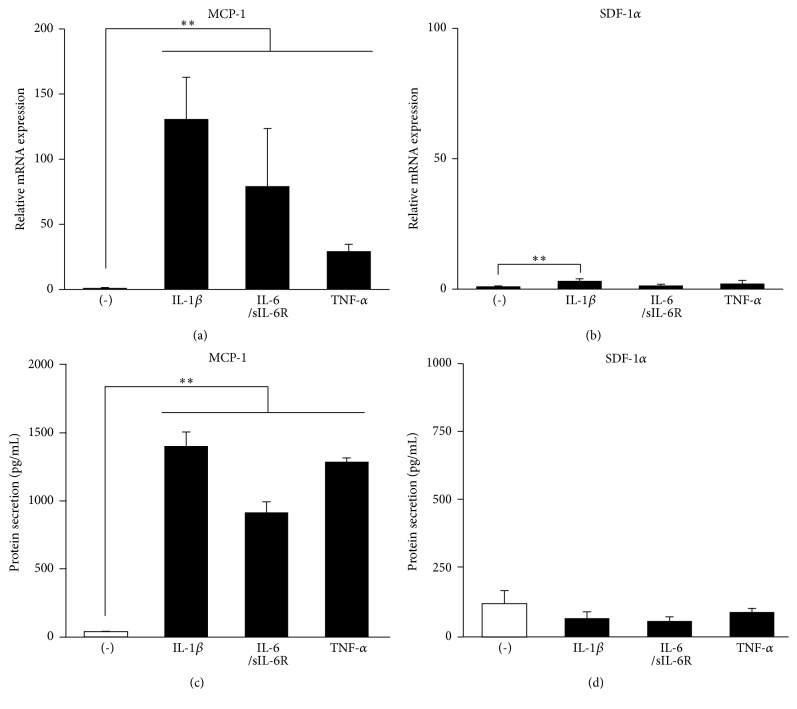
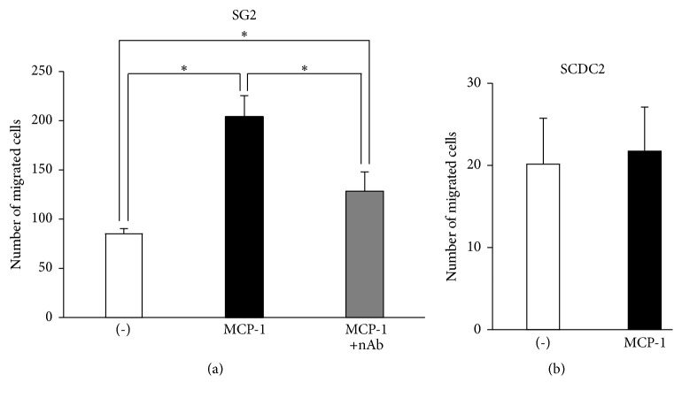
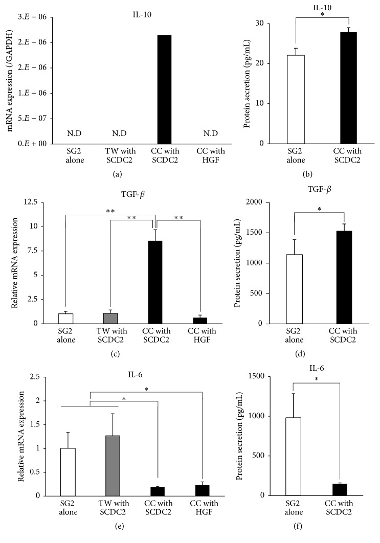
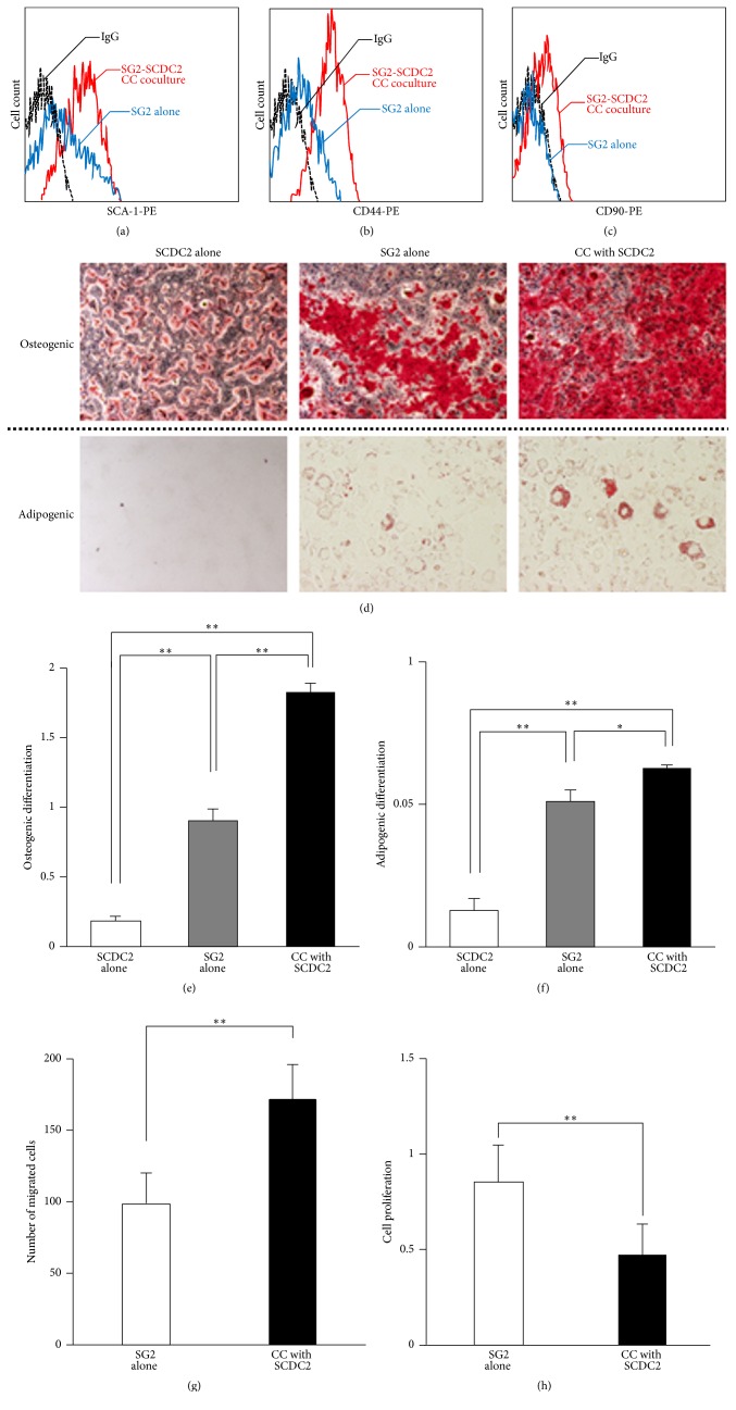
Mesenchymal stem cells (MSCs) are involved in anti-inflammatory events and tissue repair; these functions are activated by their migration or homing to inflammatory tissues in response to various chemokines. However, the mechanism by which MSCs interact with other cell types in inflammatory tissue remains unclear. We investigated the role of periodontal ligament fibroblasts (PDL-Fs) in regulating the anti-inflammatory and osteogenic abilities of bone marrow-derived- (BM-) MSCs. The expression of monocyte chemotactic protein- (MCP-)1 was significantly enhanced by stimulation of PDL-Fs with inflammatory cytokines. MCP-1 induced the migratory ability of BM-MSCs but not PDL-Fs. Expression levels of anti-inflammatory and inflammatory cytokines were increased and decreased, respectively, by direct-contact coculture between MSCs and PDL-Fs. In addition, the direct-contact coculture enhanced the expression of MSC markers that play important roles in the self-renewal and maintenance of multipotency of MSCs, which in turn induced the osteogenic ability of the cells. These results suggest that MCP-1 induces the migration and homing of BM-MSCs into the PDL inflammatory tissue. The subsequent adherence of MSCs to PDL-Fs plays an immunomodulatory role to terminate inflammation during wound healing and upregulates the expression stem cell markers to enhance the stemness of MSCs, thereby facilitating bone formation in damaged PDL tissue.


Conflict of interest statement

The authors declare that they have no competing interests.

Figures

Figure 1
Figure 1
Inflammatory cytokines promote MCP-1 production in SCDC2 cells. SCDC2 cells were stimulated with or without 10 ng/mL of IL-1β, IL-6/sIL-6R, or TNF-α. (a, b) mRNA expression levels were investigated by RT-qPCR using rat-specific primers. Reported values are normalized to Gapdh expression. The results are expressed as the fold change relative to the respective control (-). (c, d) The amount of secreted chemokines was measured using sandwich ELISA kits for rat-specific MCP-1 and SDF-1α. Data are presented as the mean ± standard deviation. P < 0.05 and ∗∗P < 0.01 as compared with the unstimulated control (-).
Figure 2
Figure 2
MCP-1 specifically induces the migration of SG2 cells rather than that of SCDC2 cells. Transwell migration assay for SG-2 cells (a) and SCDC2 cells (b) by stimulation with 10 ng/mL of MCP-1. Anti-MCP-1 neutralizing antibody (nAb) was also added with MCP-1 for migration in SG2 cells (a). The cells that migrated to the underside of the membrane were labeled with DAPI and counted under a fluorescence microscope in five high-power fields. Data are presented as the mean ± standard deviation. P < 0.05 as compared with the unstimulated control (-) for each cell line.
Figure 3
Figure 3
Expression levels of the anti-inflammatory cytokines IL-10 and TGF-β in SG2 cells increased by direct-contact coculture between SG2 and SCDC2 cells, whereas IL-6 expression decreased. SG2 cells were cocultured with SCDC2 cells or HGFs in a direct (CC) coculture system and an indirect transwell (TW) coculture system. (a, c, e) mRNA expression levels were investigated by RT-qPCR using rat-specific primers. Reported values are normalized to Gapdh expression. The results are expressed as the fold change relative to the respective control (SG2 alone). (b, d, f) Cytokines produced from the SG2 cells were quantified using sandwich ELISA with mouse-specific antibodies that show no cross-reactivity with those of rats. Quantified target proteins are presented as the mean ± standard deviations. P < 0.05, ∗∗P < 0.01.
Figure 4
Figure 4
MSC stemness of SG2 cells is enhanced by direct coculture with SCDC2 cells. Cell-surface expression levels of SCA-1 (a), CD44 (b), and CD90 (c) were analyzed with each mouse-specific antibody in SG2 cells alone (blue), SG2 cells directly cocultured with SCDC2 cells (red), and an isotype control IgG (black) using flow cytometry. (d) SG2 cells were directly (CC) cocultured on the fixed feeder SCDC2 cells as described in Section 2. The SG2 cells were incubated in osteogenic (upper panel) or adipogenic (lower panel) induction medium. The cells were evaluated for extracellular matrix mineralization by alizarin red or lipid droplets by Oil Red staining. (e) Alizarin red was extracted with 10% cetylpyridinium chloride and absorbance was measured at 540 nm. (f) Oil Red O stain was extracted with DMSO and absorbance was measured at 540 nm. (g) The migratory ability of CC cocultured SG2 cells was investigated by a transwell migration assay. (h) The cell proliferation of CC cocultured SG2 cells was examined by a WST-1 assay. The results are expressed as the fold change relative to the respective control (SG2 alone). Data are presented as the mean ± standard deviation. P < 0.05, ∗∗P < 0.01.

Similar articles

Cited by

References

    1. Prockop D. J. Marrow stromal cells as stem cells for nonhematopoietic tissues. Science. 1997;276(5309):71–74. doi: 10.1126/science.276.5309.71. - DOI - PubMed
    1. Friedenstein A. J., Chailakhjan R. K., Lalykina K. S. The development of fibroblast colonies in monolayer cultures of guinea-pig bone marrow and spleen cells. Cell and Tissue Kinetics. 1970;3(4):393–403. - PubMed
    1. Friedenstein A. J., Petrakova K. V., Kurolesova A. I., Frolova G. P. Heterotopic of bone marrow. Analysis of precursor cells for osteogenic and hematopoietic tissues. Transplantation. 1968;6(2):230–247. doi: 10.1097/00007890-196803000-00009. - DOI - PubMed
    1. Zuk P. A., Zhu M., Ashjian P., et al. Human adipose tissue is a source of multipotent stem cells. Molecular Biology of the Cell. 2002;13(12):4279–4295. doi: 10.1091/mbc.E02-02-0105. - DOI - PMC - PubMed
    1. Zhang H., Miao Z., He Z., Yang Y., Wang Y., Feng M. The existence of epithelial-to-mesenchymal cells with the ability to support hematopoiesis in human fetal liver. Cell Biology International. 2005;29(3):213–219. doi: 10.1016/j.cellbi.2004.12.007. - DOI - PubMed

LinkOut - more resources