

Bilateral Simultaneous Nonarteritic Anterior Ischaemic Optic Neuropathy: Case Report
- PMID: 28167991
- PMCID: PMC5291059
- DOI: 10.3109/01658107.2013.817593
Bilateral Simultaneous Nonarteritic Anterior Ischaemic Optic Neuropathy: Case Report
Abstract
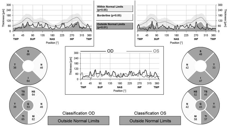
Bilateral simultaneous nonarteritic anterior ischaemic optic neuropathy (NAION) is extremely rare. A 57-year-old woman presented with bilateral optic disc oedema and peripapillary splinter haemorrhages. Initial visual acuities were hand movements in the right eye and light perception in the left eye. The patient had a mildly elevated diastolic blood pressure and glucose intolerance. Erythrocyte sedimentation rate and C-reactive protein levels were within normal limits. Temporal artery biopsy was negative for temporal arteritis. Marked visual improvement occurred in both eyes (0.8 in the right eye, 0.6 in the left eye) after systemic steroid therapy in the 16th month of follow-up.
Keywords: nonarteritic anterior ischaemic optic neuropathy treatment; simultaneous nonarteritic anterior ischaemic optic neuropathy.
Figures


References
-
- Hayreh SS, Joos KM, Podhajsky PA, Long CR. Systemic diseases associated with nonarteritic anterior ischemic optic neuropathy. Am J Ophthalmol 1994;118:766–780 - PubMed
-
- Hayreh SS. Anterior ischaemic optic neuropathy. Differentiation of arteritic from non-arteritic type and its management. Eye 1990;4:25–41 - PubMed
-
- Shibayama J, Oku H, Imamura Y, Kajiura S, Sugasawa J, Ikeda T. Bilateral, nearly simultaneous anterior ischemic optic neuropathy complicated by diabetes and bilateral, small, crowded optic discs. Jpn J Ophthalmol 2005;49:235–238 - PubMed
-
- Hayreh SS. Anterior ischemic optic neuropathy. V. Optic disc edema an early sign. Arch ophthalmol 1981;99:1030–1040 - PubMed
-
- Beri M, Klugman MR, Kohler JA, Hayreh SS. Anterior ischemic optic neuropathy. VII. Incidence of bilaterality and various influencing factors. Ophthalmology 1987;94:1020–1028 - PubMed
Publication types
LinkOut - more resources
Full Text Sources
Other Literature Sources
Research Materials