Echocardiography for patients undergoing extracorporeal cardiopulmonary resuscitation: a primer for intensive care physicians
- PMID: 28168038
- PMCID: PMC5288871
- DOI: 10.1186/s40560-017-0211-6
Echocardiography for patients undergoing extracorporeal cardiopulmonary resuscitation: a primer for intensive care physicians
Abstract
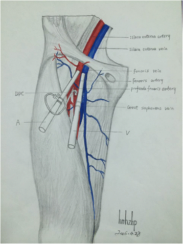
Echocardiography is an invaluable tool in the management of patients with extracorporeal cardiopulmonary resuscitation (ECPR) and subsequent extracorporeal membrane oxygenation (ECMO) support and weaning. At the very beginning, echocardiography can identify the etiology of cardiac arrest, such as massive pulmonary embolism and cardiac tamponade. Eliminating these culprits saves life and may avoid the initiation of extracorporeal cardiopulmonary resuscitation. If the underlying causes are not identified or intrinsic to the heart (e.g., such as those caused by cardiomyopathy and myocarditis), conventional cardiopulmonary resuscitation (CCPR) will continue to maintain cardiac output. The quality of CCPR can be monitored, and if cardiac output cannot be maintained, early institution of extracorporeal cardiopulmonary resuscitation may be reasonable. Cannulation is sometimes challenging for extracorporeal cardiopulmonary resuscitation patients. Fortunately, with the help of ultrasonography procedures including localization of vessels, selecting a cannula of appropriate size and confirmation of catheter tip may become easy under sophisticated hand. Monitoring of cardiac function and complications during extracorporeal membrane oxygenation support can be done with echocardiography. However, the cardiac parameters should be interpreted with understanding of hemodynamic configuration of extracorporeal membrane oxygenation. Thrombus and blood stasis can be identified with ultrasound, which may prompt mechanical and pharmacological interventions. The final step is extracorporeal membrane oxygenation weaning. A number of studies investigated the accuracy of some echocardiographic parameters in predicting success rate and demonstrated promising results. Parameters and threshold for successful weaning include aortic VTI ≥ 10 cm, LVEF > 20-25%, and lateral mitral annulus peak systolic velocity >6 cm/s. However, the effectiveness of echocardiography in ECPR patients cannot be determined in observational studies and requires randomized controlled trials in the future. The contents in this review are well known to echocardiography specialists; thus, it should be used as an educational material for emergency or intensive care physicians. There is a trend that focused echocardiography is performed by intensivists and emergency physicians.
Keywords: Cardiac arrest; Critical care; Echocardiography; Extracorporeal cardiopulmonary resuscitation; Thromboembolism.
Figures
Similar articles
-
Extracorporeal membrane oxygenation mitigates myocardial injury and improves survival in porcine model of ventricular fibrillation cardiac arrest.Scand J Trauma Resusc Emerg Med. 2019 Aug 28;27(1):82. doi: 10.1186/s13049-019-0653-z. Scand J Trauma Resusc Emerg Med. 2019. PMID: 31462264 Free PMC article.
-
RESCUE transesophageal echocardiography for monitoring of mechanical chest compressions and guidance for extracorporeal cardiopulmonary resuscitation cannulation in refractory cardiac arrest.J Clin Ultrasound. 2020 Mar;48(3):184-187. doi: 10.1002/jcu.22788. Epub 2019 Dec 10. J Clin Ultrasound. 2020. PMID: 31820822
-
Cardiac extracorporeal life support: state of the art in 2007.Cardiol Young. 2007 Sep;17 Suppl 2:104-15. doi: 10.1017/S1047951107001217. Cardiol Young. 2007. PMID: 18039404 Review.
-
Comparison of extracorporeal and conventional cardiopulmonary resuscitation: a retrospective propensity score matched study.Crit Care. 2019 Jan 28;23(1):27. doi: 10.1186/s13054-019-2320-1. Crit Care. 2019. PMID: 30691512 Free PMC article.
-
Comparing extracorporeal cardiopulmonary resuscitation with conventional cardiopulmonary resuscitation: A meta-analysis.Resuscitation. 2016 Jun;103:106-116. doi: 10.1016/j.resuscitation.2016.01.019. Epub 2016 Feb 2. Resuscitation. 2016. PMID: 26851058 Review.
Cited by
-
Recommendations for extracorporeal cardiopulmonary resuscitation (eCPR): consensus statement of DGIIN, DGK, DGTHG, DGfK, DGNI, DGAI, DIVI and GRC.Clin Res Cardiol. 2019 May;108(5):455-464. doi: 10.1007/s00392-018-1366-4. Epub 2018 Sep 4. Clin Res Cardiol. 2019. PMID: 30361819 Review.
-
Non-invasive assessment of left ventricular contractility by myocardial work index in veno-arterial membrane oxygenation patients: rationale and design of the MIX-ECMO multicentre observational study.Front Cardiovasc Med. 2024 May 28;11:1399874. doi: 10.3389/fcvm.2024.1399874. eCollection 2024. Front Cardiovasc Med. 2024. PMID: 38863897 Free PMC article.
-
Mechanical Circulatory Support for Massive Pulmonary Embolism.J Am Heart Assoc. 2025 Jan 7;14(1):e036101. doi: 10.1161/JAHA.124.036101. Epub 2024 Dec 24. J Am Heart Assoc. 2025. PMID: 39719427 Free PMC article. Review.
-
[Recommendations for extracorporeal cardiopulmonary resuscitation (eCPR) : Consensus statement of DGIIN, DGK, DGTHG, DGfK, DGNI, DGAI, DIVI and GRC].Med Klin Intensivmed Notfmed. 2018 Sep;113(6):478-486. doi: 10.1007/s00063-018-0452-8. Med Klin Intensivmed Notfmed. 2018. PMID: 29967938 German.
-
Early post-traumatic pulmonary-embolism in patients requiring ICU admission: more complicated than we think!J Thorac Dis. 2018 Nov;10(Suppl 33):S3850-S3854. doi: 10.21037/jtd.2018.09.49. J Thorac Dis. 2018. PMID: 30631496 Free PMC article. No abstract available.
References
-
- Gräsner J-T, Bossaert L. Epidemiology and management of cardiac arrest: what registries are revealing. Best Pract Res Clin Anaesthesiol. Elsevier; 2013;27:293–306 - PubMed
-
- Nürnberger A, Sterz F, Malzer R, Warenits A, Girsa M, Stöckl M, et al. Out of hospital cardiac arrest in Vienna: incidence and outcome. Resuscitation. Elsevier; 2013;84:42–7 - PubMed
-
- Rossano JW, Naim MY, Nadkarni VM, Berg RA. Epidemiology of pediatric cardiac arrest. pediatric and congenital cardiology, cardiac surgery and intensive care. London: Springer London; 2013. pp. 1275–1287.
-
- Travers AH, Perkins GD, Berg RA, Castren M, Considine J, Escalante R, et al. Part 3: adult basic life support and automated external defibrillation: 2015 International Consensus on Cardiopulmonary Resuscitation and Emergency Cardiovascular Care Science With Treatment Recommendations. Circulation. 2015;132(16 Suppl 1):S51–83. - PubMed
Publication types
LinkOut - more resources
Full Text Sources
Other Literature Sources

