. 2017:2017:8740353.
doi: 10.1155/2017/8740353. Epub 2017 Jan 12.

Right Hemisphere Remapping of Naming Functions Depends on Lesion Size and Location in Poststroke Aphasia

Affiliations

Right Hemisphere Remapping of Naming Functions Depends on Lesion Size and Location in Poststroke Aphasia

Laura M Skipper-Kallal et al. Neural Plast. 2017.

Abstract

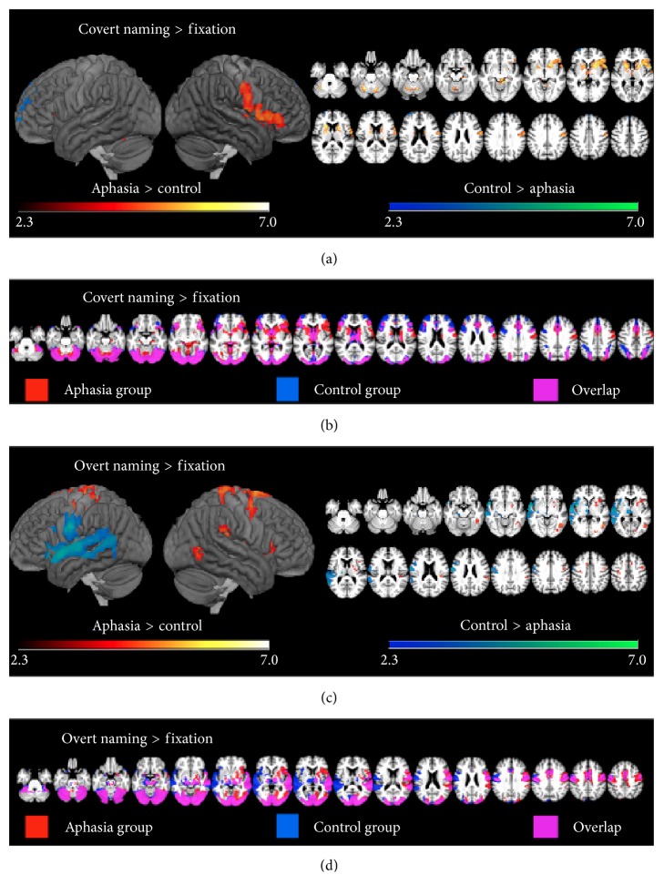
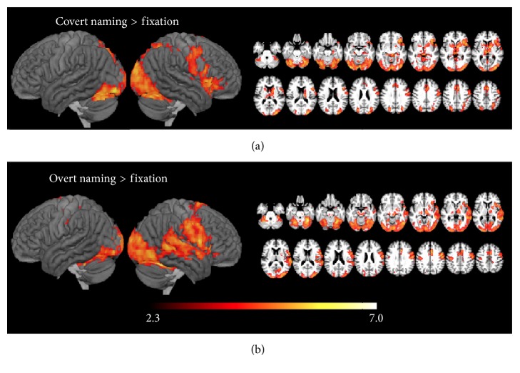
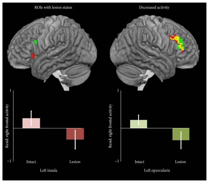
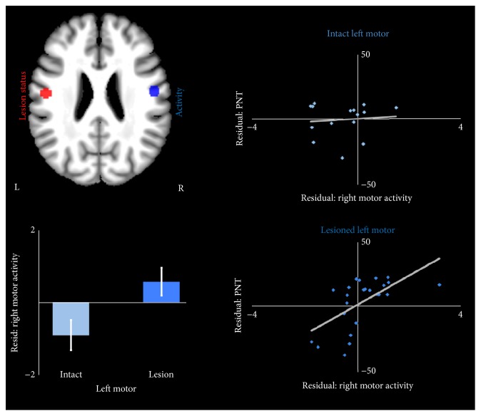
The study of language network plasticity following left hemisphere stroke is foundational to the understanding of aphasia recovery and neural plasticity in general. Damage in different language nodes may influence whether local plasticity is possible and whether right hemisphere recruitment is beneficial. However, the relationships of both lesion size and location to patterns of remapping are poorly understood. In the context of a picture naming fMRI task, we tested whether lesion size and location relate to activity in surviving left hemisphere language nodes, as well as homotopic activity in the right hemisphere during covert name retrieval and overt name production. We found that lesion size was positively associated with greater right hemisphere activity during both phases of naming, a pattern that has frequently been suggested but has not previously been clearly demonstrated. During overt naming, lesions in the inferior frontal gyrus led to deactivation of contralateral frontal areas, while lesions in motor cortex led to increased right motor cortex activity. Furthermore, increased right motor activity related to better naming performance only when left motor cortex was lesioned, suggesting compensatory takeover of speech or language function by the homotopic node. These findings demonstrate that reorganization of language function, and the degree to which reorganization facilitates aphasia recovery, is dependent on the size and site of the lesion.


Conflict of interest statement

The authors declare that there is no conflict of interests regarding the publication of this paper.

Figures

Figure 1
Figure 1
(a) Between-group contrasts showing activity during covert naming. (b) Within-group contrasts showing activity for both the aphasia and control groups during covert naming. (c) Between-group contrasts showing activity during overt naming. (d) Within-group contrasts showing activity for both the aphasia and control groups during overt naming.
Figure 2
Figure 2
Regions where large left hemisphere lesions were related to greater activity during (a) covert naming and (b) overt naming.
Figure 3
Figure 3
Lesions in the left insula and left opercularis were associated with less activity in the right middle frontal gyrus and right pars triangularis. Bar graphs show activity level in the right ROIs relative to the control sample, controlling for lesion volume.
Figure 4
Figure 4
Lesions in the left motor cortex were associated with greater activity in the right motor cortex. The bar graph shows activity level in the right motor ROI relative to controls, controlling for lesion volume. Scatter plots show the relationship between activity and performance on the PNT, controlling for lesion volume, in the intact left motor group and in the group with lesions in left motor cortex.

Similar articles

Cited by

References

    1. Pedersen P. M., Jørgensen H. S., Nakayama H., Raaschou H. O., Olsen T. S. Aphasia in acute stroke: incidence, determinants, and recovery. Annals of Neurology. 1995;38(4):659–666. doi: 10.1002/ana.410380416. - DOI - PubMed
    1. Dickey L., Kagan A., Lindsay M. P., Fang J., Rowland A., Black S. Incidence and profile of inpatient stroke-induced aphasia in Ontario, Canada. Archives of Physical Medicine and Rehabilitation. 2010;91(2):196–202. doi: 10.1016/j.apmr.2009.09.020. - DOI - PubMed
    1. Gottesman R. F., Hillis A. E. Predictors and assessment of cognitive dysfunction resulting from ischaemic stroke. The Lancet Neurology. 2010;9(9):895–905. doi: 10.1016/S1474-4422(10)70164-2. - DOI - PMC - PubMed
    1. Turkeltaub P. E., Messing S., Norise C., Hamilton R. H. Are networks for residual language function and recovery consistent across aphasic patients? Neurology. 2011;76(20):1726–1734. doi: 10.1212/WNL.0b013e31821a44c1. - DOI - PMC - PubMed
    1. Heiss W.-D., Thiel A., Kessler J., Herholz K. Disturbance and recovery of language function: correlates in PET activation studies. NeuroImage. 2003;20(supplement 1):S42–S49. doi: 10.1016/j.neuroimage.2003.09.005. - DOI - PubMed

LinkOut - more resources