. 2017 Feb 2:5:e2960.
doi: 10.7717/peerj.2960. eCollection 2017.

Medial knee joint contact force in the intact limb during walking in recently ambulatory service members with unilateral limb loss: a cross-sectional study

Affiliations

Medial knee joint contact force in the intact limb during walking in recently ambulatory service members with unilateral limb loss: a cross-sectional study

Ross H Miller et al. PeerJ. .

Abstract

Background: Individuals with unilateral lower limb amputation have a high risk of developing knee osteoarthritis (OA) in their intact limb as they age. This risk may be related to joint loading experienced earlier in life. We hypothesized that loading during walking would be greater in the intact limb of young US military service members with limb loss than in controls with no limb loss.

Methods: Cross-sectional instrumented gait analysis at self-selected walking speeds with a limb loss group (N = 10, age 27 ± 5 years, 170 ± 36 days since last surgery) including five service members with transtibial limb loss and five with transfemoral limb loss, all walking independently with their first prosthesis for approximately two months. Controls (N = 10, age 30 ± 4 years) were service members with no overt demographical risk factors for knee OA. 3D inverse dynamics modeling was performed to calculate joint moments and medial knee joint contact forces (JCF) were calculated using a reduction-based musculoskeletal modeling method and expressed relative to body weight (BW).

Results: Peak JCF and maximum JCF loading rate were significantly greater in limb loss (184% BW, 2,469% BW/s) vs. controls (157% BW, 1,985% BW/s), with large effect sizes. Results were robust to probabilistic perturbations to the knee model parameters.

Discussion: Assuming these data are reflective of joint loading experienced in daily life, they support a "mechanical overloading" hypothesis for the risk of developing knee OA in the intact limb of limb loss subjects. Examination of the evolution of gait mechanics, joint loading, and joint health over time, as well as interventions to reduce load or strengthen the ability of the joint to withstand loads, is warranted.

Keywords: Gait; Load; Military; Osteoarthritis; Prosthesis; Transfemoral; Transtibial.


Conflict of interest statement

The authors declare there are no competing interests.

Figures
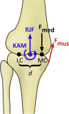
Figure 1
Figure 1. Schematic of the knee model in the frontal plane for calculating the medial knee joint contact force (Fmed).
KAM, knee adduction moment; RJF, resultant axial joint force; Fmus, muscle force, determined by the knee flexion moment; LC and MC, medial and lateral contact points, separated by distance d. Fmed is calculated by balancing the moments produced about the point LC (Schipplein & Andriacchi, 1991).
Figure 2
Figure 2. Medial knee joint contact forces in percent bodyweight (BW) during the stride, beginning at heel-strike.
Solid, dashed, and dash-dotted lines are means for control, transtibial, and transfemoral subjects. The shaded areas are ±one between-subjects standard deviation for the control subjects.
Figure 3
Figure 3. Calculated muscle forces for the quadriceps (Quads, A), hamstrings (Hams, B), and gastrocnemius (Gastroc, C) muscles during the stride, beginning at heel-strike.
Solid, dashed, and dash-dotted lines are means for control, transtibial, and transfemoral subjects. The shaded areas are ±one between-subjects standard deviation for the control subjects. Scaling factors were bodyweight (BW). The black bars along the top of each panel denote the fraction(s) of the gait cycle when this muscle group is “on” according to normative electromyograms (Sutherland, 2001), which are similar for the intact limb in limb loss subjects (Seyedali et al., 2012).
Figure 4
Figure 4. Means ± one between-subjects standard deviation for peak (A), loading rate (B), and impulse (C) of the medial knee joint contact force for the transtibial subjects (TTA), the transfemoral subjects (TFA), and the control subjects.
The limb loss bars (LL) are data for the TTA and TFA subjects combined. *, greater than control, with a large effect size (d > 0.80).
Figure 5
Figure 5. Monte Carlo simulation results for knee model parameter perturbations.
The vertical axis shows the fraction of iterations for which the medial joint contact force outcome variable was significantly greater in the limb loss group vs. the control group. The results using the original (unperturbed) parameters are not included here.
Figure 6
Figure 6. Knee flexion moment (KFM, A), knee adduction moment (KAM, B), resultant joint force along the long axis of the shank (RJF, C), and knee flexion angle (D) during the stride, beginning at heel-strike.
Solid, dashed, and dash-dotted lines are means for control, transtibial, and transfemoral subjects. The shaded areas are ±one between-subjects standard deviation for the control subjects. Scaling factors were bodyweight (BW) and height (ht).

References

    1. Amin S, Lueopongsak N, McGibbon CA, LaValley MP, Krebs DE, Felson DT. Knee adduction moment and development of chronic knee pain in elders. Arthritis & Rheumatism. 2004;51(3):371–376. doi: 10.1002/art.20396. - DOI - PubMed
    1. Andriacchi TP, Mündermann A. The role of ambulatory mechanics in the initiation and progression of knee osteoarthritis. Current Opinion in Rheumatology. 2006;18:514–518. doi: 10.1097/01.bor.0000240365.16842.4e. - DOI - PubMed
    1. Arnold EM, Ward SR, Lieber RL, Delp SL. A model of the lower limb for analysis of human movement. Annals of Biomedical Engineering. 2010;38(2):269–279. doi: 10.1007/s10439-009-9852-5. - DOI - PMC - PubMed
    1. Barrios J, Willson J. Minimum detectable change in medial tibiofemoral contact force parameters: derivation and application to a load-altering intervention. Journal of Applied Biomechanics. 2016 doi: 10.1123/jab.2016-0163. Epub ahead of print Nov 11 2016. - DOI - PubMed
    1. Bell AL, Brand RA, Peterson DR. Prediction of hip joint centre location from external landmarks. Human Movement Science. 1989;8(1):3–16. doi: 10.1016/0167-9457(89)90020-1. - DOI

LinkOut - more resources