Review
. 2017 Jan 23:4:157.
doi: 10.3389/fcell.2016.00157. eCollection 2016.

Diversity of Cnidarian Muscles: Function, Anatomy, Development and Regeneration

Affiliations
Review

Diversity of Cnidarian Muscles: Function, Anatomy, Development and Regeneration

Lucas Leclère et al. Front Cell Dev Biol. .

Abstract

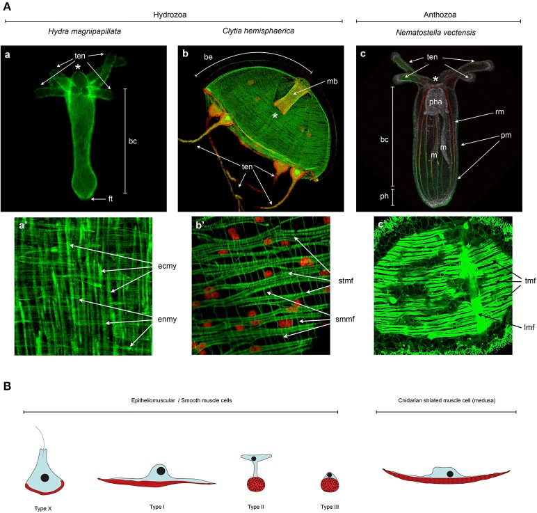
The ability to perform muscle contractions is one of the most important and distinctive features of eumetazoans. As the sister group to bilaterians, cnidarians (sea anemones, corals, jellyfish, and hydroids) hold an informative phylogenetic position for understanding muscle evolution. Here, we review current knowledge on muscle function, diversity, development, regeneration and evolution in cnidarians. Cnidarian muscles are involved in various activities, such as feeding, escape, locomotion and defense, in close association with the nervous system. This variety is reflected in the large diversity of muscle organizations found in Cnidaria. Smooth epithelial muscle is thought to be the most common type, and is inferred to be the ancestral muscle type for Cnidaria, while striated muscle fibers and non-epithelial myocytes would have been convergently acquired within Cnidaria. Current knowledge of cnidarian muscle development and its regeneration is limited. While orthologs of myogenic regulatory factors such as MyoD have yet to be found in cnidarian genomes, striated muscle formation potentially involves well-conserved myogenic genes, such as twist and mef2. Although satellite cells have yet to be identified in cnidarians, muscle plasticity (e.g., de- and re-differentiation, fiber repolarization) in a regenerative context and its potential role during regeneration has started to be addressed in a few cnidarian systems. The development of novel tools to study those organisms has created new opportunities to investigate in depth the development and regeneration of cnidarian muscle cells and how they contribute to the regenerative process.

Keywords: cnidaria; development; epitheliomuscular cells; evolution; muscle; myoepithelial cells; regeneration.


Figures

Figure 1
Figure 1
Bilaterian and cnidarian phyolgenies. (A) Metazoan phylogeny, highlighting the pivotal position of cnidarians as the sister group to extant bilaterian animals. The position of Ctenophora and Porifera (sponges) outside the Bilateria remains controversial (as indicated by dashed lines). (B) Cnidarian phylogeny showing the relationships between the main lineages based on recently published data (Chang et al., ; Zapata et al., 2015).
Figure 2
Figure 2
Cnidarian life cycles. The life cycles of (A) the solitary fresh water polyp Hydra, (B) the marine jellyfish Clytia (both hydrozoans) and (C) the anthozoan polyp Nematostella. At the lower part of the panels are indicated their asexual reproductive potentials (budding, physal pinching) that give rise to new (A) Hydra or (C) Nematostella polyps, or (B) juvenile Clytia medusae, respectively. Under harsh environmental conditions, gonads develop and sexual reproduction in (A) Hydra can occur. Depending on the species, Hydra can be gonochoric or hermaphroditic (represented here). After fertilization, embryonic development occurs within a solid capsule that, after hatching, frees a juvenile Hydra. (B) Clytia and (C) Nematostella are gonochoric and oocytes and sperm are released into the water column. After fertilization, embryonic development leads to the formation of swimming planula larvae that after metamorphosis develop into (B) a polyp colony for Clytia or (C) a solitary juvenile polyp for Nematostella.
Figure 3
Figure 3
Cnidarian muscle functions. (A) Planula larva crawling, (B), Hydra polyp somersaulting, (C) jellyfish pulsation, (D) guided tentacle retraction of the jellyfish to bring the food toward the mouth, (E) digestive peristaltic movements of the polyp (red rings along the body column indicate circumferential muscle contractions), (F) protective retraction of the polyp in response to predation pressure.
Figure 4
Figure 4
Cnidarian muscle diversity. (A) Muscle networks of (a,a') Hydra magnipapillata, (b,b') Clytia hemisphaerica jellyfish and (c,c') Nematostella vectensis. The upper panels show the muscle network of entire organisms and the lower panel magnification of certain body regions to highlight the orientation and fine structure of the muscles. (A) Living Lifeact:GFP transgene (Seybold et al., 2016) labeling actin filaments, (c) fixed MyHC1::mCherry (Renfer et al., 2010) transgene labeling the actin fibers of the retractor muscles, co-labeled with phalloidin. All other panels (a',b,b',c') are phalloidin stainings. Image labels are as follows: (*) mouth, (ten) tentacles, (bc) body column, (ft) foot, (be) bell, (mb) manubrium, (pha) pharynx, (m) mesentery, (ph) physa, (rm) retractor muscle, (pm) parietal muscle, (ecmy) ectodermal myonemes, (enmy) endodermal myonemes, (stmf) striated muscle fibers, (smmf) smooth-like muscle fibers, (tmf) transversal muscle fibers, (lmf) loongitudinal muscle fibers. (a,a') Image courtesy of Aufschnaiter and Hobmayer, (b,b') images from Kraus et al. (2015) and (c,c') Image from Amiel et al. (2015) as well as courtesy of Amiel. (B) Epitheliomuscular cell type diversity in Cnidaria. After Krasińska (1914) and Doumenc (1979) in Seipel and Schmid (2006), and Jahnel et al. (2014).
Figure 5
Figure 5
Cnidarian “muscle” gene repertoire. Overview of the cnidarian “muscle” gene repertoire in regard to known bilaterian myogenic factors. Cnidarians are represented by Hydra, Clytia, Podocoryna, and Nematostella. The potential role in myogenesis of a given gene in the indicated species has been assessed by functional studies if available or by published gene expression patterns, (n/a) no information available. References cited in this figure: (1) Chapman et al., ; (2) Hoffmann and Kroiher, ; (3) Jager et al., ; (4) Steinmetz et al., ; (5) Chiori et al., ; (6) Kraus et al., ; (7) Stierwald et al., ; (8) Spring et al., ; (9) Galle et al., ; (10) Spring et al., ; (11) Ryan et al., ; (12) Matus et al., ; (13) Saina and Technau, ; (14) Putnam et al., ; (15) Magie et al., ; (16) Nakanishi et al., ; (17) Genikhovich and Technau, ; (18) Martindale et al., ; (19) Ryan et al., ; (20) Ryan et al., .
Figure 6
Figure 6
Cnidarian regeneration potential. Regenerative capacities of (A) Hydra, (B) Clytia medusa and (D) Nematostella as cnidarian representatives. (C) Illustrates the re-symmetrization process of juvenile medusa that is not regeneration per se, but allows a quick regain of the medusa functionality. (E) Illustrates the transdifferentiation and regeneration potential of striated muscle cells isolated from jellyfish and cultured in vitro. See text for further details.

Similar articles

Cited by

References

    1. Abrams M. J., Basinger T., Yuan W., Guo C.-L., Goentoro L. (2015). Self-repairing symmetry in jellyfish through mechanically driven reorganization. Proc. Natl. Acad. Sci. U.S.A. 112, E3365–E3373. 10.1073/pnas.1502497112 - DOI - PMC - PubMed
    1. Adams J. C., Brancaccio A. (2015). The evolution of the dystroglycan complex, a major mediator of muscle integrity. Biol. Open 4, 1163–1179. 10.1242/bio.012468 - DOI - PMC - PubMed
    1. Adell T., Saló E., Boutros M., Bartscherer K. (2009). Smed-Evi/Wntless is required for beta-catenin-dependent and -independent processes during planarian regeneration. Development 136, 905–910. 10.1242/dev.033761 - DOI - PubMed
    1. Aerne B., Gröger H., Schuchert P., Spring J., Schmid V. (1996). The polyp and its medusa: a molecular approach. Sci. Mar. 60, 7–16.
    1. Alberts B., Johnson A., Lewis J., Morgan D., Raff M., Roberts K., et al. (2015). Molecular Biology of the Cell, 6th Edn. New York, NY: Garland Science.