. 2017 Jan 16;4(2):106-118.
doi: 10.1002/acn3.384. eCollection 2017 Feb.

White matter predicts functional connectivity in premanifest Huntington's disease

Affiliations

White matter predicts functional connectivity in premanifest Huntington's disease

Peter McColgan et al. Ann Clin Transl Neurol. .

Abstract

Objectives: The distribution of pathology in neurodegenerative disease can be predicted by the organizational characteristics of white matter in healthy brains. However, we have very little evidence for the impact these pathological changes have on brain function. Understanding any such link between structure and function is critical for understanding how underlying brain pathology influences the progressive behavioral changes associated with neurodegeneration. Here, we demonstrate such a link between structure and function in individuals with premanifest Huntington's.

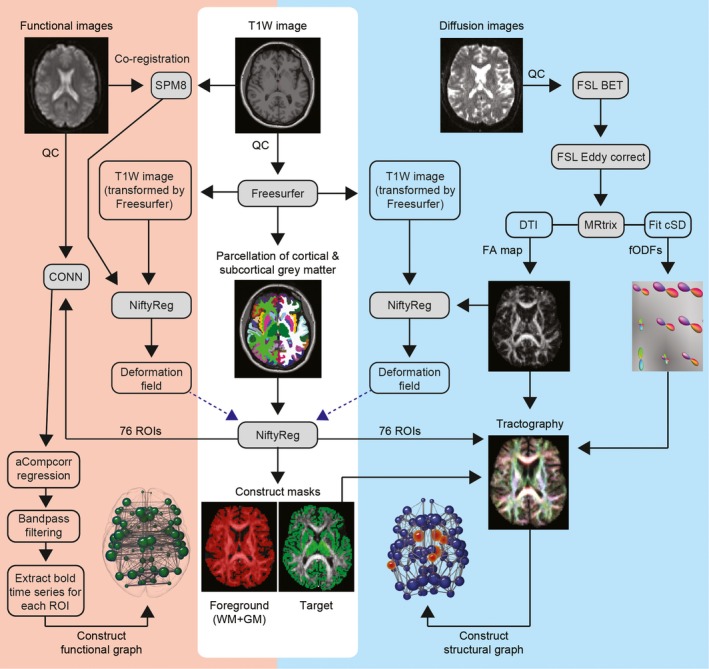
Methods: Using diffusion tractography and resting state functional magnetic resonance imaging to characterize white matter organization and functional connectivity, we investigate whether characteristic patterns of white matter organization in the healthy human brain shape the changes in functional coupling between brain regions in premanifest Huntington's disease.

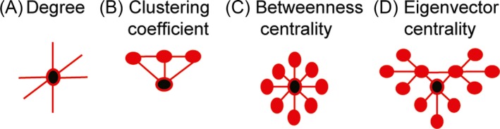
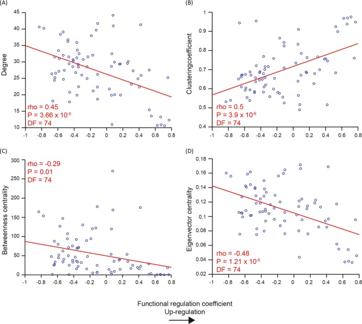
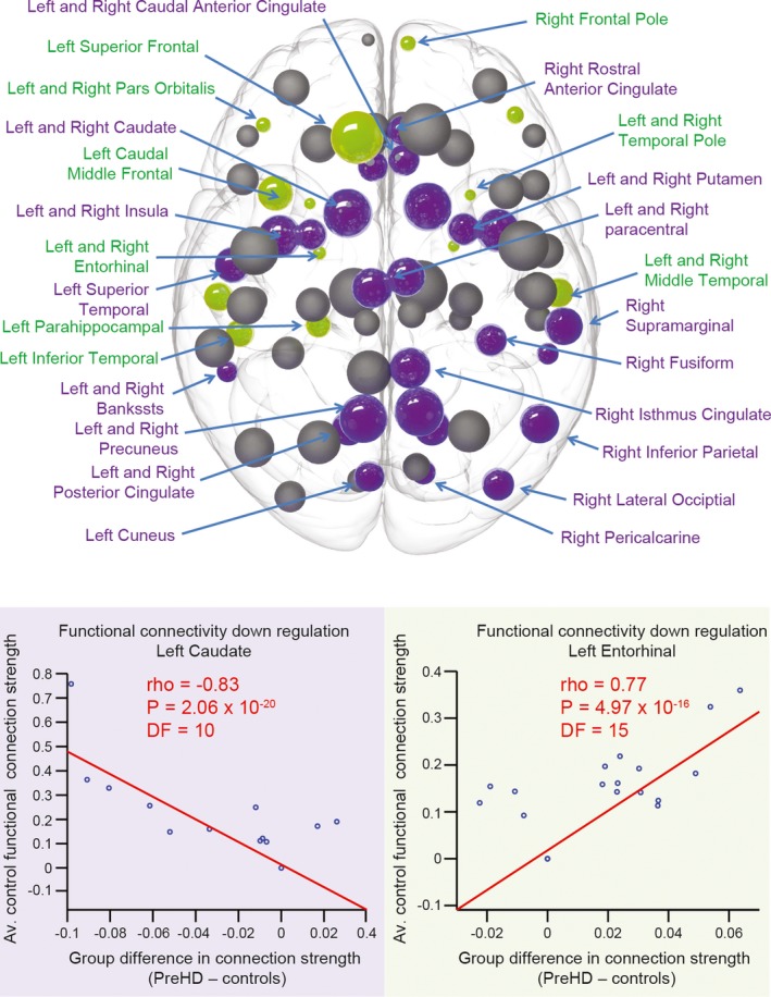
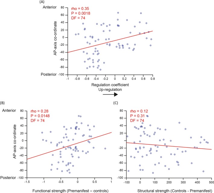
Results: We find changes in functional connectivity in premanifest Huntington's disease that link directly to underlying patterns of white matter organization in healthy brains. Specifically, brain areas with strong structural connectivity show decreases in functional connectivity in premanifest Huntington's disease relative to controls, while regions with weak structural connectivity show increases in functional connectivity. Furthermore, we identify a pattern of dissociation in the strongest functional connections between anterior and posterior brain regions such that anterior functional connectivity increases in strength in premanifest Huntington's disease, while posterior functional connectivity decreases.

Interpretation: Our findings demonstrate that organizational principles of white matter underlie changes in functional connectivity in premanifest Huntington's disease. Furthermore, we demonstrate functional antero-posterior dissociation that is in keeping with the caudo-rostral gradient of striatal pathology in HD.


Figures

Figure 1
Figure 1
Resting state fMRI and diffusion tractography processing pipelines. BET, brain extraction tool; CONN, functional connectivity toolbox; CSD, constrained spherical deconvolution; DTI, diffusion tensor imaging; FA, fractional anisotropy; fODF, fibre orientation distribution function; GM, gray matter; QC, quality control; WM, white matter; SPM, statistical parametric mapping.
Figure 2
Figure 2
Schematic description of graph theory metrics. (A) Degree is the number of connections a brain region has. (B) Clustering coefficient indicates how highly connected a region is to its neighbors and (C) Betweenness centrality represents brain region network traffic. (D) Eigenvector centrality represents network traffic along the brains ‘busiest’ pathways. Black circles represent regions with high degree, clustering coefficient, betweenness centrality or eigenvector centrality. These graph theory metrics correspond to the graph metrics on the y‐axis of Figures 2, 3 and 4.
Figure 3
Figure 3
Prediction of functional upregulation based on healthy white matter organization. Regions with (A) low degree, (B) high clustering and (C,D) low network traffic (betweenness and eigenvector centrality) show greatest functional upregulation in pre‐HD. The graph theory metric value of a brain region in the average control WM brain network, on the y‐axis, is plotted against the functional regulation coefficient for that corresponding brain region, on the x‐axis. The red line represents a least squares linear regression line. rho, correlation coefficient; DF, degrees of freedom; WM, white matter.
Figure 4
Figure 4
Functional regulation analysis. For each brain region in the average control network, correlations were performed against the strength of functional connection to all other 75 regions in the network (where a functional connection was present) and average group differences (pre‐HD minus controls) in these functional connections. Upregulation is defined as a positive correlation (stronger control connections show greater increases in pre‐HD), whereas downregulation is defined as a negative correlation (stronger control connections show greater decreases in pre‐HD). Brain regions that show significant positive (green) and negative (purple) correlations are highlighted. The size of the sphere represents the number of structural connections (thus largest spheres indicate hub brain regions). Correlation plots showing the brain regions with the most significant positive (green) and negative (purple) correlations are also displayed below. For each plot each data point represents a connection to the brain region specified. The strength of that connection for the avaerage control network, on the y‐axis, is plotted against the difference (pre‐HD minus controls) of that connection's strength on the x‐axis. The red line represents a least squares linear regression line. rho, correlation coefficient; DF, degrees of freedom.
Figure 5
Figure 5
A–P correlation analysis for functional regulation, functional and structural strength. More anterior regions show greater increases in (A) regulation coefficient and (B) functional but not (C) structural strength in pre‐HD. Each data point represents a brain region. The coordinate of that brain region along the anterior–posterior axis, on the y‐axis, is plotted against regulation coefficient or strength on the x‐axis.The red line represents a least squares linear regression line. rho, correlation coefficient; DF, degrees of freedom.

Similar articles

Cited by

References

    1. Ross CA, Aylward EH, Wild EJ, et al. Huntington disease: natural history, biomarkers and prospects for therapeutics. Nat Rev Neurol 2014;10:204–216. - PubMed
    1. McColgan P, Seunarine KK, Razi A, et al. Selective vulnerability of rich club brain regions is an organizational principle of structural connectivity loss in Huntington's disease. Brain 2015;138(Pt 11):3327–44. - PMC - PubMed
    1. Zhou J, Gennatas ED, Kramer JH, et al. Predicting regional neurodegeneration from the healthy brain functional connectome. Neuron 2012;73:1216–1227. - PMC - PubMed
    1. Tabrizi SJ, Reilmann R, Roos RA, et al. Potential endpoints for clinical trials in premanifest and early Huntington's disease in the TRACK‐HD study: analysis of 24 month observational data. Lancet Neurol 2012;11:42–53. - PubMed
    1. Kloppel S, Gregory S, Scheller E, et al. Compensation in preclinical Huntington's disease: evidence from the track‐on HD study. EBioMedicine 2015;2:1420–1429. - PMC - PubMed

LinkOut - more resources