. 2017 Apr 20;45(7):3888-3905.
doi: 10.1093/nar/gkx081.

Dynamic assembly of Hda and the sliding clamp in the regulation of replication licensing

Affiliations

Dynamic assembly of Hda and the sliding clamp in the regulation of replication licensing

Jin S Kim et al. Nucleic Acids Res. .

Abstract

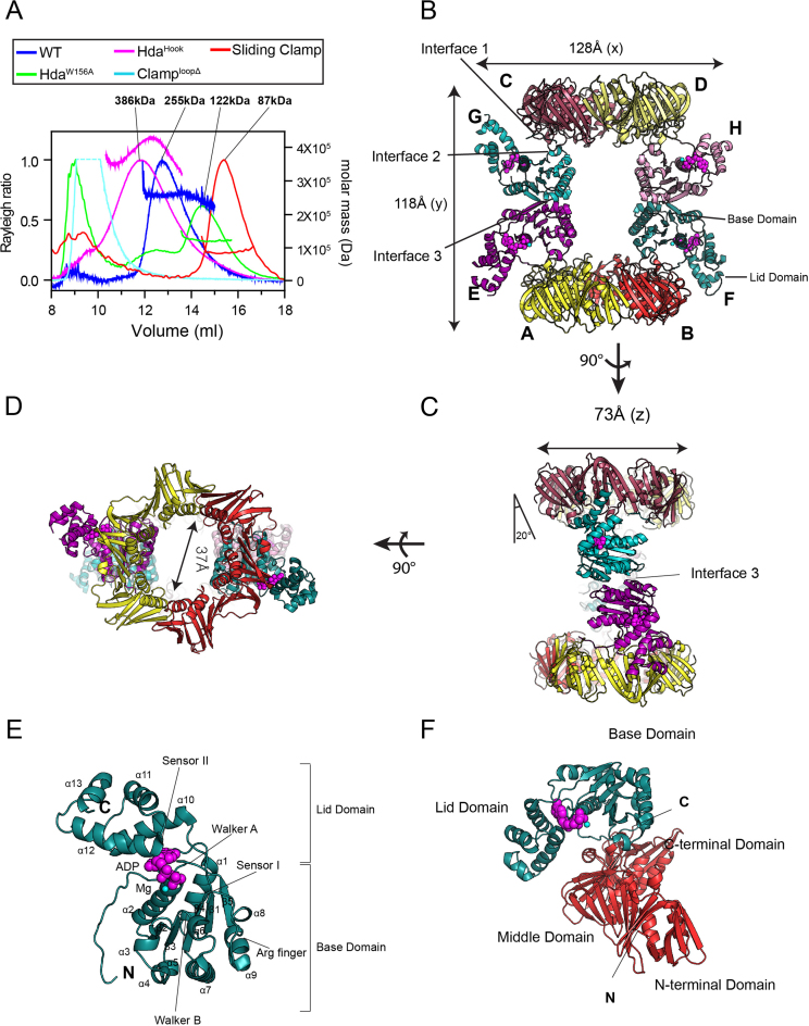
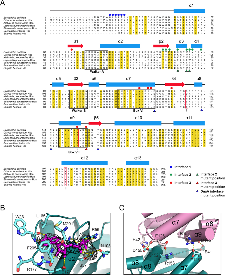
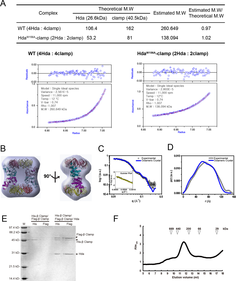
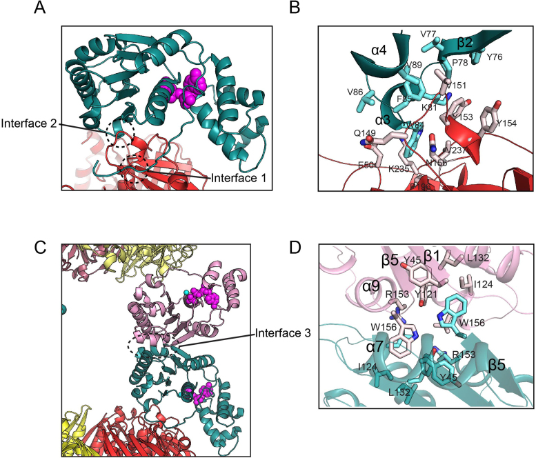
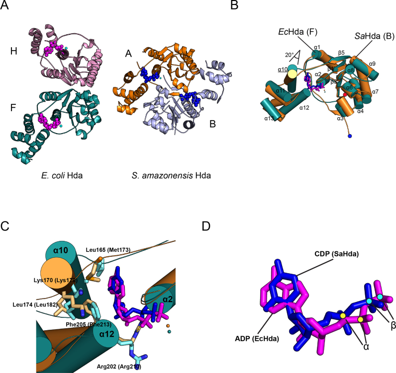
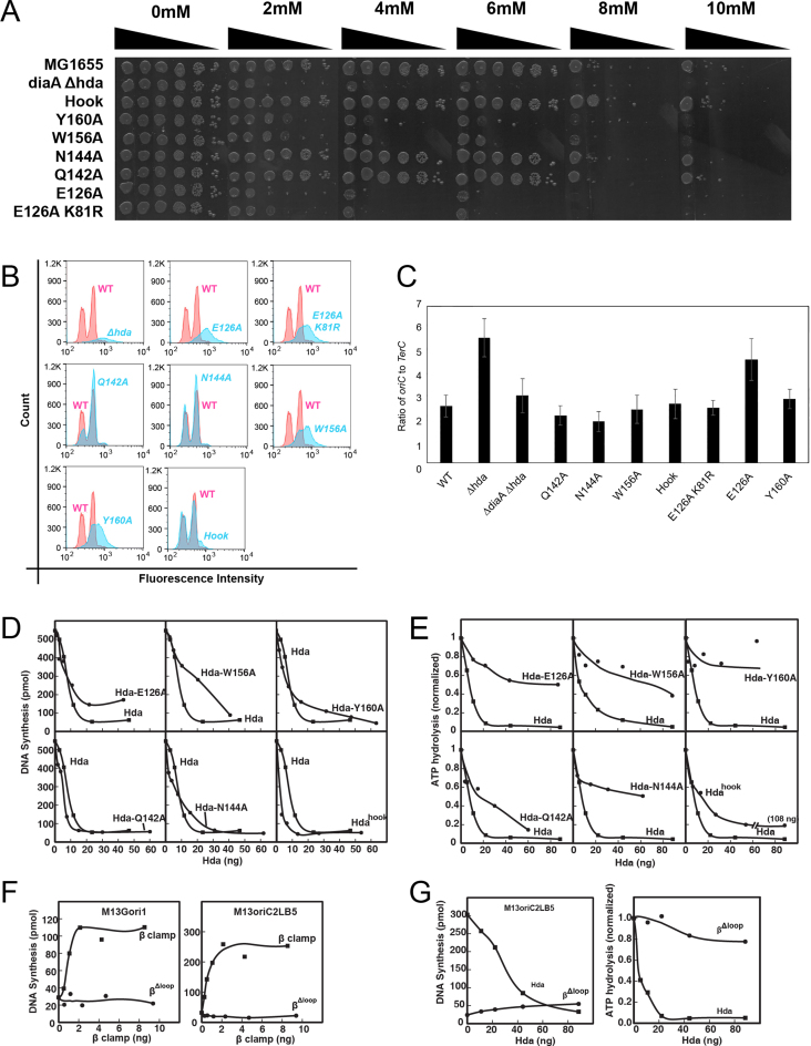
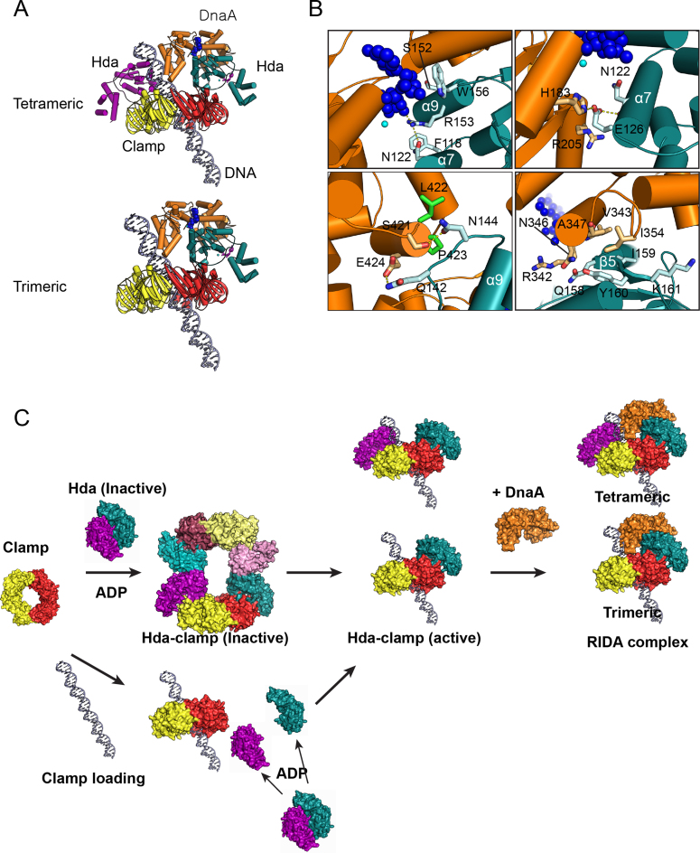
Regulatory inactivation of DnaA (RIDA) is one of the major regulatory mechanisms of prokaryotic replication licensing. In RIDA, the Hda-sliding clamp complex loaded onto DNA directly interacts with adenosine triphosphate (ATP)-bound DnaA and stimulates the hydrolysis of ATP to inactivate DnaA. A prediction is that the activity of Hda is tightly controlled to ensure that replication initiation occurs only once per cell cycle. Here, we determined the crystal structure of the Hda-β clamp complex. This complex contains two pairs of Hda dimers sandwiched between two β clamp rings to form an octamer that is stabilized by three discrete interfaces. Two separate surfaces of Hda make contact with the β clamp, which is essential for Hda function in RIDA. The third interface between Hda monomers occludes the active site arginine finger, blocking its access to DnaA. Taken together, our structural and mutational analyses of the Hda-β clamp complex indicate that the interaction of the β clamp with Hda controls the ability of Hda to interact with DnaA. In the octameric Hda-β clamp complex, the inability of Hda to interact with DnaA is a novel mechanism that may regulate Hda function.


Figures

Figure 1.
Figure 1.
Structure of the Hda–β clamp complex. (A) SEC-MALS analysis of the Hda–β clamp complexes containing wild-type Hda or the indicated mutants, and of the β clamp alone. (B) Structure of the Hda–β clamp octameric complex in ribbon representation. Each β clamp is labeled A, B, C or D. Each Hda molecule is labeled E, F, G or H. ADP (magenta) and magnesium ion (cyan) in each Hda protomer are shown as spheres. Two orthogonal views of the Hda–β clamp complex structure in (B) are shown in (C) and (D). Interface 1, 2 and 3 are indicated in panels B and C. (E) Close-up view of the Hda molecule (chain F). The secondary structures, AAA+ conserved motifs and N- and C-termini of Hda are marked. (F) Heterodimeric arrangement of Hda (chain F) and the β clamp (chain B).
Figure 2.
Figure 2.
Secondary structure and key structural features of Hda (A) Sequence alignment of Hda from several bacterial species: Escherichia coli, Citrobacter rodentium, Klebsiella pneumoniae, Legionella pneumophila, Shewanella amazonensis, Salmonella enterica and Shigella flexneri. The secondary structural elements are shown at the top. The residues of interface 1, 2 and 3 are marked in blue, green or red dots, respectively. The substituted residues of the mutants used in SEC-MALS analyses are indicated by green and red triangles, and the residues mutated for DnaA binding are indicated by blue triangles. The conserved Walker A, Walker B, Box VI and Box VII motifs are denoted by black boxes. Sensor I, sensor II and arginine finger are shown in red boxes with I, II and R characters below, respectively. (B) The nucleotide-binding site of Hda (chain F). Residues surrounding ADP (magenta), Mg2+ ion (cyan dot) and a water molecule (red dot) are indicated. The electron density shown as a black mesh is a 2mFo-DFc simulated-annealing omit-map contoured at 1σ for ADP, Mg and the water molecule. Hydrogen bonds between ADP and surrounding residues are represented by yellow dots. (C) Close-up view of interface 3 that also shows the arginine finger in the E and G chains of Hda (purple and teal).
Figure 3.
Figure 3.
Analytical ultracentrifugation and SAXS analysis of the Hda–β clamp complex. (A) Sedimentation equilibrium data were evaluated using a nonlinear least-squares curve-fitting algorithm for the Hda–β clamp complex containing wild-type (WT) Hda or the W156A mutant. The expected sedimentation behavior for the Hda–β clamp complex is shown as a red line in comparison with the experimental data (blue circles). (B–D) SAXS analysis of the WT Hda–β clamp complex is shown. Blue envelopes are superimposed with the Hda–β clamp complex in two different orientations (B). The SAXS profile of the solution structure and the pair distance distribution functions [p(r)] for the Hda–β clamp complex are shown in (C) and (D). The experimental data are indicated by open circles, and the data calculated from the crystal structure are shown as solid blue lines. (E) Pull-down analyses of the His-β clamp and the Flag-β clamp in the absence or presence of Hda, as indicated. The lanes labeled ‘His’ contain the eluate from the Ni2+-NTA bead pull-down. Likewise, lanes labeled ‘Flag’ contain samples from the subsequent anti-Flag pull-down of that same eluate. Positions of the Flag-β clamp, His-β clamp and Hda and molecular weight markers are indicated. (F) SEC profile of the Hda–β clamp complex at low concentration (100 μg/ml, 20 μg). The elution position of molecular weight markers are also shown.
Figure 4.
Figure 4.
Three interfaces of the Hda–β clamp complex. (A) Interface 1 and 2 formed between Hda (Chain F; deep teal) and the β clamp (Chain B; red) are circled. (B) Close-up view of the interface 2. (C) Interface 3 is formed between the two Hda protomers. (D) Close-up view of the interface 3.
Figure 5.
Figure 5.
Comparison of the structures of the Escherichia coli Hda dimer with the Shewanella amazonensis Hda dimer. (A) The E. coli Hda dimer (chain F, teal; chain H, pink) in the Hda–β clamp complex is shown. Nucleotides are shown as pink spheres (left). The S. amazonensis Hda dimer (chain A, light blue; chain B, orange) is also shown. Nucleotides are shown in blue. The F chain of EcHda and the A chain of SaHda are displayed in the same orientation. (B) The superimposed structures of EcHda (chain F, teal) of the Hda–β clamp complex and SaHda (3BOS; chain A, orange) together with secondary structural elements are shown. ADP in EcHda and CDP in SaHda are shown in magenta and blue sticks, respectively. Each N-terminus of EcHda or SaHda is marked at the bottom with red or blue dots, respectively. (C) Close-up view of the nucleotide-binding site of the superimposed structures shows the interactions between nucleotide and hydrophobic residues of the lid domain. (D) The superimposed ADP and CDP of EcHda and SaHda, and the α-phosphate and β-phosphate marked by yellow or cyan dots are shown. The sticks are colored as in (B).
Figure 6.
Figure 6.
In vivo and In vitro analyses of Hda mutants (A) In vivo test of sensitivity of Escherichia coli mutants to hydroxyurea. After serial dilution, the sensitivity of E. coli strains encoding mutant Hdas to the indicated concentrations of HU was measured as described in ‘Materials and Methods’ section. Cultures were normalized for cell density based on OD600nm prior to serial dilutions. (B) Flow cytometry analysis of strains expressing hda alleles. Cells (50 000) from the indicated strains were analyzed after staining with PicoGreen. Chromosome equivalents were determined using E. coli MG1655 as the control and shown in red in comparison with isogenic strains bearing the indicated hda allele shown in blue. Fluorescence intensity (abscissa) is presented in logarithmic scale. (C) Ratio of oriC to TerC of the strain encoding WT or mutant hda. (D) DNA synthesis was measured in reactions containing a supercoiled plasmid bearing oriC in the presence of WT Hda or mutants as described in ‘Materials and Methods’ section. (E) Hydrolysis of ATP bound to DnaA by WT Hda or mutants. (F) DNA synthesis was measured in reactions containing M13Gori1 ssDNA (left) or M13oriC2LB5 supercoiled DNA (right), other reaction components and increasing amounts of WT β or the βΔloop clamp mutant followed by incubation at 30°C for 10 or 20 min as described in ‘Materials and Methods’ section. (G) Influence of the βΔloop clamp mutant on the activity of DnaA in the RIDA assay that measures DNA replication of an oriC-containing plasmid (left) and ATP hydrolysis (right).
Figure 7.
Figure 7.
Structural basis for RIDA. (A) Models of the complex containing the β clamp, Hda and DnaA bound to DNA. The model was built by aligning Hda (from the tetrameric or trimeric Hda–clamp complex) onto the DnaA dimer (generated from SWISS-MODEL). The chains of the EcHda–β clamp tetramer are represented by the same colors as in the octameric complex of Figure 1B; EcDnaA is colored in orange. (B) The panels display the close-up views of the interfaces between EcDnaA and EcHda. (C) RIDA activation model. Dimeric Hda forms a complex with the β clamp to form an inactive octamer. In the clamp loading process, the octameric complex dissociates and assembles on DNA as a trimeric or tetrameric complex. One possible mechanism involves one Hda molecule remaining bound to a protomer of the β clamp, which is loaded onto the DNA to form the trimer (not shown). Alternatively, Hda monomers bind to the β clamp already loaded on DNA to form a trimeric or tetrameric complex. To bind ATP-DnaA and to stimulate ATP hydrolysis, part of Hda may be released from the loop (residues 148–154) at interface 2.

References

    1. Skarstad K., Katayama T.. Regulating DNA replication in bacteria. Cold Spring Harb. Perspect. Biol. 2013; 5:a012922. - PMC - PubMed
    1. Riber L., Frimodt-Møller J., Charbon G., Løbner-Olesen A.. Multiple DNA binding proteins contribute to timing of chromosome replication in E. coli. Front. Mol. Biosci. 2016; 3:29. - PMC - PubMed
    1. Duderstadt K.E., Chuang K., Berger J.M.. DNA stretching by bacterial initiators promotes replication origin opening. Nature. 2011; 478:209–213. - PMC - PubMed
    1. Kaguni J.M. DnaA: controlling the initiation of bacterial DNA replication and more. Annu. Rev. Microbiol. 2006; 60:351–375. - PubMed
    1. Sutton M.D., Carr K.M., Vicente M., Kaguni J.M.. Escherichia coli DnaA protein. The N-terminal domain and loading of DnaB helicase at the E. coli chromosomal origin. J. Biol. Chem. 1998; 273:34255–34262. - PubMed

MeSH terms

LinkOut - more resources