Observational Study
. 2017 Sep;27(9):3593-3599.
doi: 10.1007/s00330-017-4761-8. Epub 2017 Feb 6.

High b-value diffusion-weighted imaging in progressive multifocal leukoencephalopathy in HIV patients

Affiliations
Observational Study

High b-value diffusion-weighted imaging in progressive multifocal leukoencephalopathy in HIV patients

Claudia Godi et al. Eur Radiol. 2017 Sep.

Abstract

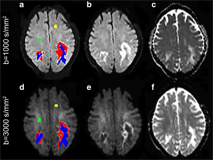
Objectives: An ill-defined hyperintense edge and hypointense core on diffusion-weighted imaging (DWI) is typical of progressive multifocal leukoencephalopathy (PML). We aimed to investigate whether a b-value of 3,000 s/mm2 (b3000) can improve visualisation of PML, or provide different structural information compared to 1,000 s/mm2 (b1000).

Methods: We retrospectively identified HIV-positive patients with confirmed PML studied under a clinical protocol including both b1000 and b3000 DWI. The rim and core of each PML lesion and normal-appearing white matter (NAWM) were outlined on trace-weighted DWI. Signal intensities, apparent diffusion coefficient (ADC) values and volumes were measured and compared between b1000 and b3000.

Results: Nine lesions from seven patients were analysed. The rim and core were better visualised on b3000, with higher signal of the rim and lower signal of the core compared to NAWM. The hyperintense rim had non-restricted average ADCs, but included foci of low ADC on both b3000 and b1000. Despite similar total lesion volumes, b3000 displayed significantly larger core and smaller rim volumes than b1000.

Conclusion: b3000 improves visualisation of this important PML hallmark. Moreover, b3000 partly reclassifies tissue from rim into core, and might provide potentially more accurate biomarkers of PML activity and prognosis.

Key points: • B3000 improves contrast resolution between lesion rim, core and normal-appearing white matter. • B3000 improves identification of the typical rim-and-core pattern of PML lesions. • B3000 and b1000 similarly identify lesions, but b3000 results in smaller rims and larger cores. • B3000 excludes some high diffusion components from rim, reclassifying them into core. • B3000 DWI may provide more precise PML biomarkers of disease activity and tissue damage.

Keywords: Diffusion MRI; Diffusion magnetic resonance imaging; Diffusion-weighted MRI; Human immunodeficiency virus; Progressive multifocal leukoencephalopathy.


Figures

Fig. 1
Fig. 1
b1000 and b3000 volumes (in mm3) of the rim and core of all progressive multifocal leukoencephalopathy (PML) lesions are shown in panels A and B, respectively. Overall, the rim volumes on b3000 were lower, and the core volumes higher than their counterparts on b1000 diffusion-weighted imaging without any significant change in the total lesion volume being noted (C)
Fig. 2
Fig. 2
A representative patient is shown in images AF. On b1000 (B) and b3000 (E) trace-weighted diffusion-weighted imaging, progressive multifocal leukoencephalopathy (PML) rim is outlined in red, PML core in blue, and normal-appearing white matter (NAWM) in yellow and green (A, D). Although similar lesion volumes were identified by the two b-values (sum of red and blue components), the rim in b1000 appeared thicker than in b3000 (B, E); conversely, the core in b3000 was more identifiable and larger than in b1000, not only on trace-weighted images (B, E) but also on apparent diffusion coefficient (ADC) maps (C, F)
Fig. 3
Fig. 3
At b1000 the hyperintense progressive multifocal leukoencephalopathy (PML) rim is a heterogeneous region that corresponds to intermediate apparent diffusion coefficient (ADC) values on average, although some spots of low ADC values within the rim are observed (arrowheads). However the rim on b3000 diffusion-weighted imaging (DWI) contains a higher proportion of low ADC values (arrows), suggesting that the more stringent delineation of the lesion rim on b3000 results in exclusion of some areas with increased diffusion

References

    1. Ke A, El M, Ep R. Progressive multifocal leuko-encephalopathy; a hitherto unrecognized complication of chronic lymphatic leukaemia and Hodgkin’s disease. Brain. 1958;81:93–111. doi: 10.1093/brain/81.1.93. - DOI - PubMed
    1. Zurhein G, Sm C. Particles resembling papova viruses in human cerebral demyelinating disease. Science. 1965;148:1477–1479. doi: 10.1126/science.148.3676.1477. - DOI - PubMed
    1. Padgett BL, Walker DL, ZuRhein GM, et al. Cultivation of papova-like virus from human brain with progressive multifocal leucoencephalopathy. Lancet. 1971;1:1257–1260. doi: 10.1016/S0140-6736(71)91777-6. - DOI - PubMed
    1. Holman RC, Török TJ, Belay ED, et al. Progressive multifocal leukoencephalopathy in the United States, 1979–1994: increased mortality associated with HIV infection. Neuroepidemiology. 1998;17:303–309. doi: 10.1159/000026184. - DOI - PubMed
    1. Kleinschmidt-DeMasters BK, Tyler KL. Progressive multifocal leukoencephalopathy complicating treatment with natalizumab and interferon beta-1a for multiple sclerosis. N Engl J Med. 2005;353:369–374. doi: 10.1056/NEJMoa051782. - DOI - PubMed

Publication types