Review
. 2017 Jun;22(2):93-108.
doi: 10.1007/s10911-017-9372-0. Epub 2017 Feb 6.

The Terminal End Bud: the Little Engine that Could

Affiliations
Review

The Terminal End Bud: the Little Engine that Could

Ingrid S Paine et al. J Mammary Gland Biol Neoplasia. 2017 Jun.

Abstract

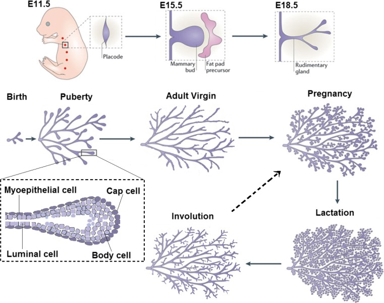
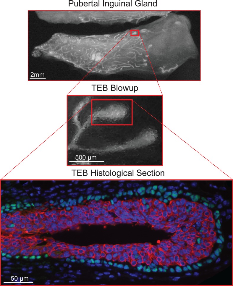
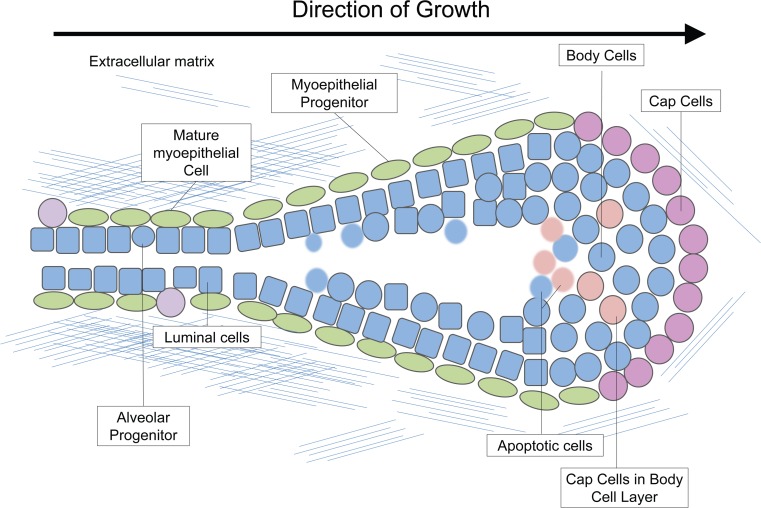
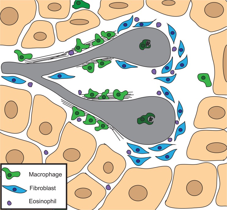
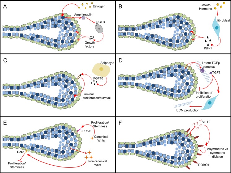
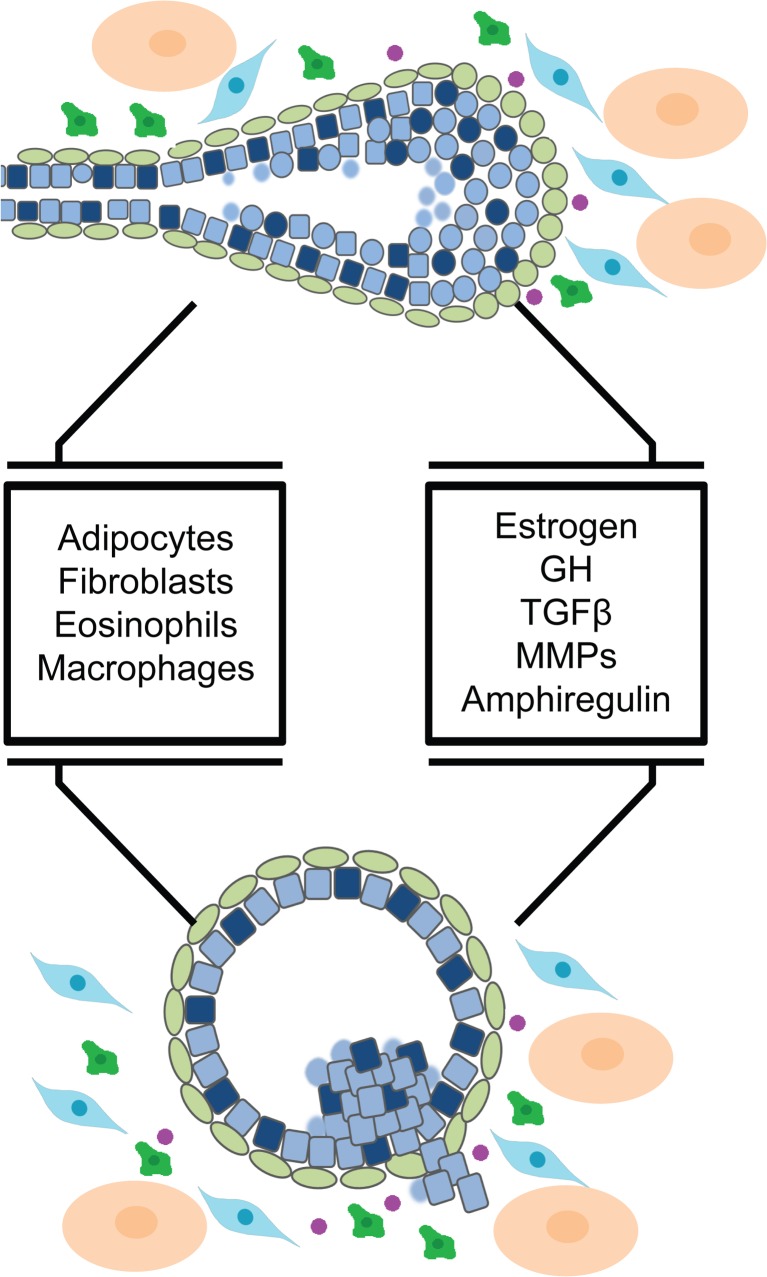
The mammary gland is one of the most regenerative organs in the body, with the majority of development occurring postnatally and in the adult mammal. Formation of the ductal tree is orchestrated by a specialized structure called the terminal end bud (TEB). The TEB is responsible for the production of mature cell types leading to the elongation of the subtending duct. The TEB is also the regulatory control point for basement membrane deposition, branching, angiogenesis, and pattern formation. While the hormonal control of TEB growth is well characterized, the local regulatory factors are less well understood. Recent studies of pubertal outgrowth and ductal elongation have yielded surprising details in regards to ongoing processes in the TEB. Here we summarize the current understanding of TEB biology, discuss areas of future study, and discuss the use of the TEB as a model for the study of breast cancer.

Keywords: Cap cell; Ductal elongation; Mammary gland; Terminal end bud.


Conflict of interest statement

This work was supported by National Science Foundation grant DMS-126375. Michael T. Lewis is a Limited Partner in StemMed Ltd. and a Manager in StemMed Holdings L.L.C.

Figures

Fig. 1
Fig. 1
Overview of Mammary Development. Mammary placodes are formed on E11.5. The mammary bud sinks into the fat pad precursor on E15.5 and expands to form a rudimentary duct by E18.5. The gland remains relatively quiescent until the onset of puberty, when terminal end buds are formed and direct ductal elongation (blow up of pubertal time point). During pregnancy alveolar budding and differentiation take place to give rise to the milk producing cells. Adapted from [146]
Fig. 2
Fig. 2
Terminal End Bud Whole Mounts and Histology. During puberty, terminal end buds direct the growth of the duct through the fat pad and are seen at the leading edge of the growing duct (top panel). The terminal end buds are bulb shaped structures that undergo regular bifurcation events (middle panel). A histological section of a terminal end bud reveals two compartments that can be distinguished by expression of cell surface markers (bottom panel)
Fig. 3
Fig. 3
Terminal End Bud. The terminal end bud is a bulbous structure made up of two main compartments known as the cap and body cell layers. The cells in the front of the terminal end bud (pink) are least differentiated (cap cells and body cells), whereas cells become more differentiated in the narrowing region (myoepithelial progenitors-green) and the neck of the terminal end bud (luminal cells- blue, mature myoepithelial cells-green). Some cap cells can be found in the body cell layer (beige). The extra-cellular matrix is light around the leading tip of the terminal end bud and becomes a complex meshwork in the neck of the terminal end bud. Apoptosis in the body cell layer contributes to lumen formation (faded blue and beige)
Fig. 4
Fig. 4
Stromal Cell Types Involved in Ductal Elongation. The mammary stroma contains many cell types that together are instructive for ductal morphogenesis. Three cell types are localized around active terminal end buds and are important for ductal elongation. Fibroblasts (blue) are found around the advancing tip of the terminal end bud and produce extracellular matrix proteins and growth factors. Macrophages (green) are found at the neck of the terminal end bud where they organize extracellular matrix proteins into a complex meshwork. Macrophages are also found within the body cell layer where they help to remove apoptotic bodies. Eosinophils (purple) are recruited to terminal end buds by eotaxin and are important for branching. The stroma also contains adipocytes (beige)
Fig. 5
Fig. 5
Signaling Essential for Terminal End Buds. a Estrogen from the ovaries signals through the Estrogen Receptor positive luminal cells to produce the paracrine factor amphiregulin, which acts on stromal cells to produce additional growth factors. b Growth hormone from the pituitary acts on fibroblasts near the terminal end bud to produce insulin growth factor which promotes proliferation in the epithelial cells. c The adipocytes produce fibroblast growth factor 10 which acts specifically on basal cells to induce a pro-proliferative response in the luminal compartment. d Transforming Growth Factor Beta is produced by the epithelial cells and secreted as a latent complex. Once cleaved it has divergent effects on stromal and epithelial cell types. e Canonical and non-canonical Wnt signaling oppose each other to control stemness in the mammary gland during puberty. f SLIT2 is produced by both luminal and basal cells in the terminal end bud and binds to the ROBO receptor found only in the basal compartment to promote survival of cap cells
Fig. 6
Fig. 6
The Terminal End Bud as A Model for Breast Cancer. The terminal end bud has many important features in common with early breast cancers including close contact with specialized stromal cell types, and several signaling pathways that are important for cell growth and survival

References

    1. Veltmaat JM, et al. Identification of the mammary line in mouse by Wnt10b expression. Dev Dyn. 2004;229(2):349–356. doi: 10.1002/dvdy.10441. - DOI - PubMed
    1. Sakakura T, et al. Biology of mammary fat pad in fetal mouse: capacity to support development of various fetal epithelia in vivo. Development. 1987;100(3):421–430. - PubMed
    1. Dunbar ME, et al. Parathyroid hormone-related protein signaling is necessary for sexual dimorphism during embryonic mammary development. Development. 1999;126(16):3485–3493. - PubMed
    1. Hogg NA, Harrison CJ, Tickle C. Lumen formation in the developing mouse mammary gland. J Embryol Exp Morphol. 1983;73:39–57. - PubMed
    1. Inman JL, et al. Mammary gland development: cell fate specification, stem cells and the microenvironment. Development. 2015;142(6):1028–1042. doi: 10.1242/dev.087643. - DOI - PubMed

Publication types