

Potential therapeutic role of punicalagin against mechanical-trauma-induced stress urinary incontinence via upregulation of Nrf2 and TGF-β1 signaling : Effect of punicalagin on mechanical trauma induced SUI
- PMID: 28168411
- PMCID: PMC5437194
- DOI: 10.1007/s00192-017-3283-x
Potential therapeutic role of punicalagin against mechanical-trauma-induced stress urinary incontinence via upregulation of Nrf2 and TGF-β1 signaling : Effect of punicalagin on mechanical trauma induced SUI
Erratum in
-
Correction to: Potential therapeutic role of punicalagin against mechanical-trauma-induced stress urinary incontinence via upregulation of Nrf2 and TGF-β1 signaling : Effect of punicalagin on mechanical trauma induced SUI.Int Urogynecol J. 2024 May;35(5):1105-1106. doi: 10.1007/s00192-024-05790-8. Int Urogynecol J. 2024. PMID: 38625605 No abstract available.
Abstract
Introduction and hypothesis: We investigated the effect of punicalagin (PUN; 2,3-hexahydroxydiphenoyl-gallagyl-D-glucose), on mechanical-trauma-induced stress urinary incontinence (SUI) in mouse and the mechanisms underlying any effects.
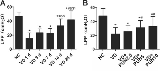
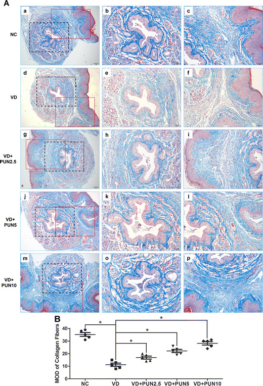
Methods: Ninety virgin female C57BL/6 mice were randomized into six groups: five groups underwent vaginal distention (VD) for 1 h and leak-point pressure (LPP) was measured on the 1st, 3rd, 7th, 14th, and 28th day following (VD groups 1 d, 3 d, 7 d, 14 d, and 28 d). The sixth group was a noninstrumented control (NC) group. Then, 75 virgin female C57BL/6 mice were randomized into five groups: a VD group (that just underwent VD) and an NC group were orally administered saline every day for 7 days; and three VD + PUN groups that underwent VD and were orally administered PUN respectively at 2.5, 5, and 10 mg/kg every day for 7 days. LPP was tested on the day 7, then all mice were sacrificed and their urethras and anterior vaginal walls harvested for Masson staining, immunohistochemistry study, Western blot analysis, and quantitative polymerase chain reaction (qPCR).
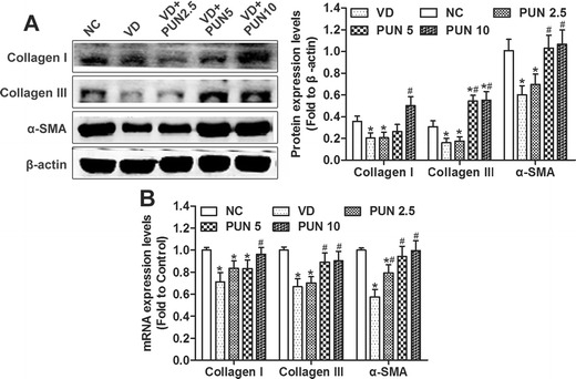
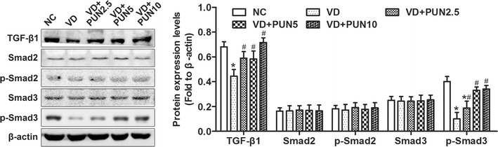
Results: LPPs after VD were significantly lower than the NC group, and the LPPs of mice on days 14 and 28 day after VD were significantly higher than on the days 1, 3, and 7. PUN significantly improved VD-induced drops in LPP and alleviated VD-induced decrease of collagen I, collagen III, α-smooth muscle actin (SMA), transforming growth factor (TGF)-β1, and p-Smad3, nuclear factor-erythroid 2 p45-related factor 2 (Nrf2), and glutathione peroxidase (GPx1) protein levels, and increase of 8-hydroxydeoxyguanosine (OHdG) in urethra and anterior vaginal wall. PUN also up-regulated the expression of manganese superoxide dismutase (MnSOD), whereas protein levels of Smad 2, p-Smad2, and Smad3 were not changed.
Conclusions: PUN exerts certain therapeutic effect on mechanical-trauma-induced SUI in mice, which might be through the activation of TGF-β1/Smad3 and Nrf2/antioxidant response element (ARE) signaling activation.
Keywords: Extracellular matrix; Mechanical trauma; Oxidative damage; Punicalagin; Stress urinary incontinence; Vaginal distension.
Conflict of interest statement
None.
Figures

Similar articles
-
Dimethyl fumarate ameliorates stress urinary incontinence by reversing ECM remodeling via the Nrf2-TGF-β1/Smad3 pathway in mice.Int Urogynecol J. 2022 May;33(5):1231-1242. doi: 10.1007/s00192-021-05061-w. Epub 2022 Jan 4. Int Urogynecol J. 2022. PMID: 34982187
-
Protective Role of Nuclear Factor Erythroid-2-Related Factor 2 against Mechanical Trauma-Induced Apoptosis in a Vaginal Distension-Induced Stress Urinary Incontinence Mouse Model.Oxid Med Cell Longev. 2019 Mar 6;2019:2039856. doi: 10.1155/2019/2039856. eCollection 2019. Oxid Med Cell Longev. 2019. PMID: 30962861 Free PMC article.
-
Mechanism of Mechanical Trauma-Induced Extracellular Matrix Remodeling of Fibroblasts in Association with Nrf2/ARE Signaling Suppression Mediating TGF-β1/Smad3 Signaling Inhibition.Oxid Med Cell Longev. 2017;2017:8524353. doi: 10.1155/2017/8524353. Epub 2017 Oct 3. Oxid Med Cell Longev. 2017. PMID: 29109834 Free PMC article.
-
Puerarin protects fibroblasts against mechanical stretching injury through Nrf2/TGF-β1 signaling pathway.Int Urogynecol J. 2022 Sep;33(9):2565-2576. doi: 10.1007/s00192-022-05325-z. Epub 2022 Aug 13. Int Urogynecol J. 2022. PMID: 35962806 Review.
-
Activation of Nrf2/AREs-mediated antioxidant signalling, and suppression of profibrotic TGF-β1/Smad3 pathway: a promising therapeutic strategy for hepatic fibrosis - A review.Life Sci. 2020 Sep 1;256:117909. doi: 10.1016/j.lfs.2020.117909. Epub 2020 Jun 5. Life Sci. 2020. PMID: 32512009 Review.
Cited by
-
Punicalagin inhibited proliferation, invasion and angiogenesis of osteosarcoma through suppression of NF‑κB signaling.Mol Med Rep. 2020 Sep;22(3):2386-2394. doi: 10.3892/mmr.2020.11304. Epub 2020 Jul 6. Mol Med Rep. 2020. PMID: 32705250 Free PMC article.
-
Dimethyl fumarate ameliorates stress urinary incontinence by reversing ECM remodeling via the Nrf2-TGF-β1/Smad3 pathway in mice.Int Urogynecol J. 2022 May;33(5):1231-1242. doi: 10.1007/s00192-021-05061-w. Epub 2022 Jan 4. Int Urogynecol J. 2022. PMID: 34982187
-
Nampt promotes fibroblast extracellular matrix degradation in stress urinary incontinence by inhibiting autophagy.Bioengineered. 2022 Jan;13(1):481-495. doi: 10.1080/21655979.2021.2009417. Bioengineered. 2022. PMID: 34967693 Free PMC article.
-
Cytokine modulation in pelvic organ prolapse and urinary incontinence: from molecular insights to therapeutic targets.Mol Med. 2024 Nov 13;30(1):214. doi: 10.1186/s10020-024-00989-3. Mol Med. 2024. PMID: 39538179 Free PMC article. Review.
-
BMMSC-sEV-derived miR-328a-3p promotes ECM remodeling of damaged urethral sphincters via the Sirt7/TGFβ signaling pathway.Stem Cell Res Ther. 2020 Jul 16;11(1):286. doi: 10.1186/s13287-020-01808-2. Stem Cell Res Ther. 2020. PMID: 32678010 Free PMC article.
References
-
- Di Biase M, Malhorta N, Kocjancic E. Management of stress urinary incontinence. Semin Colon Rectal Surg. 2016;27(1):46–50. doi: 10.1053/j.scrs.2015.12.009. - DOI
MeSH terms
Substances
LinkOut - more resources
Full Text Sources
Other Literature Sources
Medical
Miscellaneous
