. 2017 Jul;24(7):838-849.
doi: 10.1097/GME.0000000000000840.

Vaginal estrogen: a dual-edged sword in postoperative healing of the vaginal wall

Affiliations

Vaginal estrogen: a dual-edged sword in postoperative healing of the vaginal wall

Christopher M Ripperda et al. Menopause. 2017 Jul.

Abstract

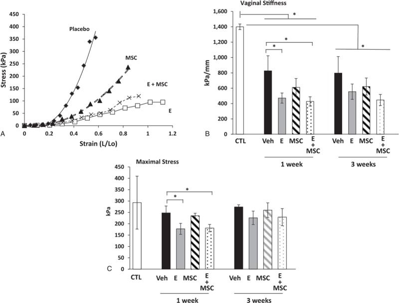
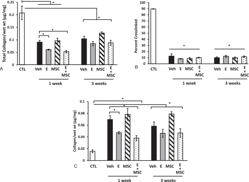
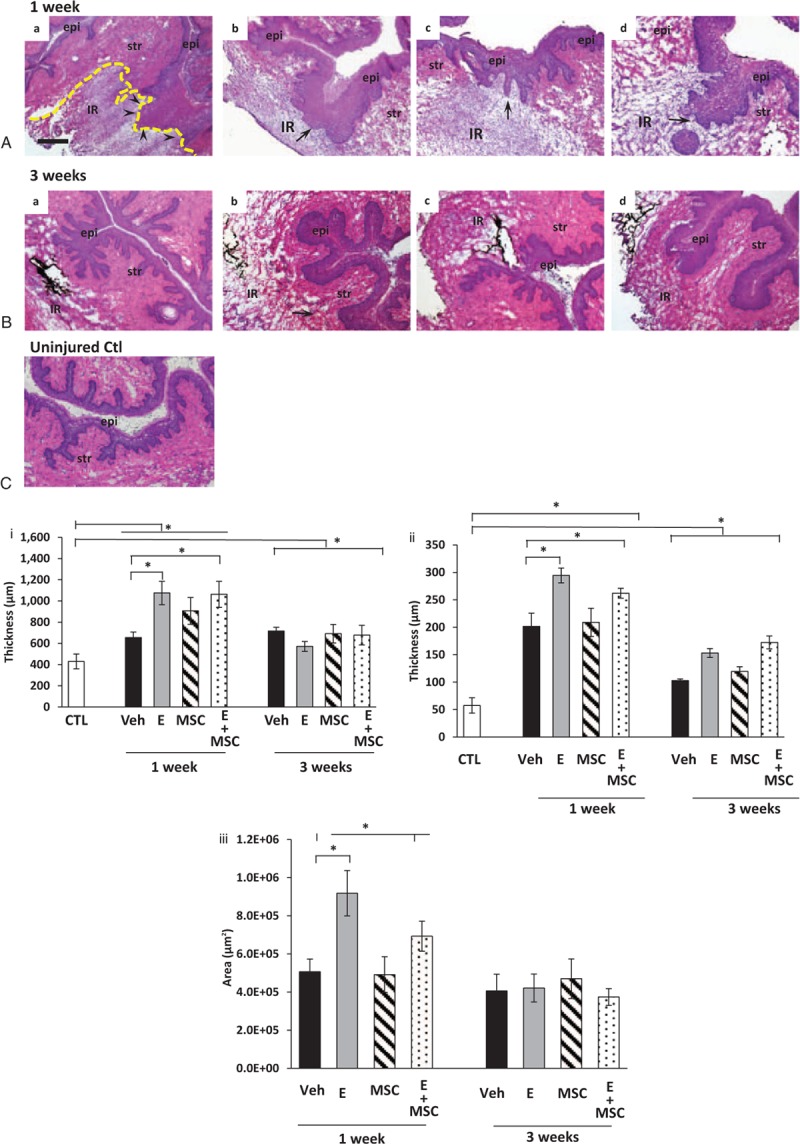
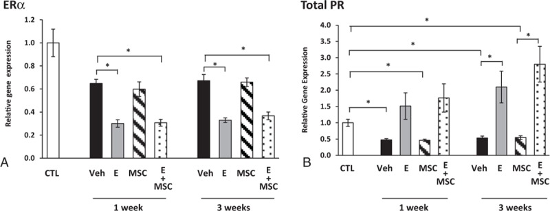
Objective: Reconstructive surgery for pelvic organ prolapse is plagued with high failure rates possibly due to impaired healing or regeneration of the vaginal wall. Here, we tested the hypothesis that postoperative administration of local estrogen, direct injection of mesenchymal stem cells (MSCs), or both lead to improved wound healing of the injured vagina in a menopausal rat model.

Methods: Ovariectomized rats underwent surgical injury to the posterior vaginal wall and were randomized to treatment with placebo (n = 41), estrogen cream (n = 47), direct injection of MSCs (n = 39), or both (n = 43).

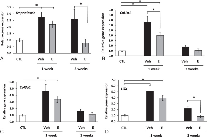
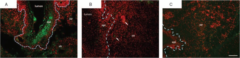
Results: MSCs did not survive after injection and had no appreciable effects on healing of the vaginal wall. Acute postoperative administration of vaginal estrogen altered the response of the vaginal wall to injury with decreased stiffness, decreased collagen content, and decreased expression of transcripts for matrix components in the stromal compartment. Conversely, vaginal estrogen resulted in marked proliferation of the epithelial layer and increased expression of genes related to epithelial barrier function and protease inhibition. Transcripts for genes involved in chronic inflammation and adaptive immunity were also down-regulated in the estrogenized epithelium.

Conclusions: Collectively, these data indicate that, in contrast to the reported positive effects of preoperative estrogen on the uninjured vagina, acute administration of postoperative vaginal estrogen has adverse effects on the early phase of healing of the stromal layer. In contrast, postoperative estrogen plays a positive role in healing of the vaginal epithelium after injury.


Conflict of interest statement

Financial disclosure/conflicts of interest: None reported.

Figures

FIG. 1
FIG. 1
Biomechanical properties of the injured vaginal wall. Stress-strain relationships (A) of vaginal tissues from ovariectomized rats treated with placebo cream/placebo injection (Placebo, diamond, n = 8), estrogen cream/placebo injection (E, open square, n = 9), placebo cream/mesenchymal stem cell (MSCs) injection (MSC, triangle, n = 9), and estrogen cream + MSC injection (E + MSC, X, n = 9). Quantification of vaginal stiffness (B) and maximum vaginal stress (C) are presented as mean ± SEM. Two-way analysis of variance. CTL, control; Veh, vehicle.
FIG. 2
FIG. 2
Effect of vaginal estrogen and mesenchymal stem cells (MSCs) on collagen content of the injured vaginal wall. (A) Total collagen content per milligram wet weight of vaginal stromal compartment collected from the site of injury 1 or 3 weeks after injury. Data represent injured vaginal stroma from ovariectomized rats treated with placebo cream/placebo injection (Placebo, solid bar), estrogen cream/placebo injection (E, gray bar, n = 9), placebo cream/MSC injection (MSC, lined bar), and estrogen cream + MSC injection (E + MSC, dotted bar). (B) Percentage of cross-linked mature collagen, and (C) newly synthesized immature collagen per milligram wet weight. Data represent mean ± SEM of 9-12 rats in each group. Two-way analysis of variance (P < 0.05). CTL, control; Veh; vehicle.
FIG. 3
FIG. 3
Histologic analysis of injury sites from ovariectomized rats treated with placebo, estrogen, MSCs, or MSCs + E at 1 (A) or 3 (B) weeks. epi, epithelium, str, stroma; IR, injury reaction. Arrows denote basement membrane at site of injury. Arrowheads in Aa indicate disruption of the basement membrane. (C) Uninjured control. Sections were stained with hematoxylin and eosin and captured at the same magnification (bar = 400 μm). Dark staining indicates incorporation of India ink at injury site. Dashed line indicates area of injury reaction. Lower panel (i), thickness of posterior vaginal stromal compartment or epithelium, (ii) at the injury site (μm) from ovariectomized rats treated with placebo cream/placebo injection (Placebo, solid bar), estrogen cream/placebo injection (E, gray bar, n = 9), placebo cream/mesenchymal stem cell injection (MSC, lined bar), or estrogen cream + MSC (E + MSC, dotted bar) 1 and 3 weeks after injury. Results were compared with uninjured untreated control ovariectomized rats (CTL). (iii) Area of injury reaction (μm2). Data represent mean ± SEM of 9-10 animals in each treatment group except uninjured controls (n = 3). Two-way analysis of variance (P < 0.05). Veh, vehicle; CTL, control.
FIG. 4
FIG. 4
Effect of vaginal estrogen and mesenchymal stem cells (MSCs) on gene expression of ERα (A) or progesterone receptor (PR) (B) in the vaginal stroma before or after injury. Relative gene expression was determined by quantitative PCR in vaginal stroma from ovariectomized rats before injury (CTL) or 1 and 3 weeks after injury treated with placebo cream/placebo injection (Placebo, solid bar), estrogen cream/placebo injection (E, gray bar, n = 9), placebo cream/MSC injection (MSC, lined bar), or estrogen cream + MSC (E + MSC, dotted bar). Data represent mean ± SEM of 7-9 animals in each group. P < 0.05 compared with uninjured controls, analysis of variance with Dunn posthoc testing. ERα, estrogen receptor alpha; CTL, control; Veh, vehicle.
FIG. 5
FIG. 5
Effect of vaginal estrogen on gene expression of tropoelastin (A), Col1a1 (B), Col3a1 (C), or lysyl oxidase (LOX, D) in vaginal stroma before or after injury. Relative gene expression was determined by qPCR. Data represent mean ± SEM of 7-9 animals in each group. P < 0.05 compared with uninjured controls, analysis of variance. qPCR, quantitative polymerase chain reaction; Col1a1, collagen type 1 a1; Col3a1, collagen type 3 a1; CTL, control; SEM, standard error of the mean; Veh, vehicle.
FIG. 6
FIG. 6
Immunostaining of green fluorescent protein (GFP)-labeled MSCs in vaginal wall of ovariectomized rats after injury. Ovariectomized rats underwent surgical injury of the posterior vaginal wall. Four days after injury, GFP-labeled mesenchymal stem cells (MSCs) were injected into the site of injury. Tissues were collected 3 hours (A), 3 days (1 week after injury) (B), or 17 days (3 weeks after injury) after injection (C). Sections were stained for GFP (green) and counterstained with Hoechst nuclear stain (red) and represent experiments conducted in duplicate. The border between epithelium and stroma is indicated by a dashed line. Arrows note MSCs in the 1-week group. epi, epithelium; str, stroma. Arrows denote basement membrane at site of injury. Bar = 100 μm.

Similar articles

Cited by

References

    1. Olsen AL, Smith VJ, Bergstrom JO, Colling JC, Clark AL. Epidemiology of surgically managed pelvic organ prolapse and urinary incontinence. Obstet Gynecol 1997; 89:501–506. - PubMed
    1. DeLancey JOL. The hidden epidemic of pelvic floor dysfunction: achievable goals for improved prevention and treatment. Am J Obstet Gynecol 2005; 192:1488–1495. - PubMed
    1. Abramov Y, Webb AR, Botros SM, Goldberg RP, Ameer GA, Sand PK. Effect of bilateral oophorectomy on wound healing of the rabbit vagina. Fertil Steril 2011; 95:1467–1470. - PubMed
    1. Balgobin S, Montoya TI, Shi H, et al. Estrogen alters remodeling of the vaginal wall after surgical injury in guinea pigs. Biol Reprod 2013; 89:138. - PMC - PubMed
    1. Montoya TI, Maldonado PA, Acevedo JF, Word RA. Effect of vaginal or systemic estrogen on dynamics of collagen assembly in the rat vaginal wall. Biol Reprod 2015; 92:43. - PMC - PubMed