. 2017 Apr 1;19(4):535-545.
doi: 10.1093/neuonc/now235.

Germline and somatic BAP1 mutations in high-grade rhabdoid meningiomas

Affiliations

Germline and somatic BAP1 mutations in high-grade rhabdoid meningiomas

Ganesh M Shankar et al. Neuro Oncol. .

Abstract

Background: Patients with meningiomas have widely divergent clinical courses. Some entirely recover following surgery alone, while others have relentless tumor recurrences. This clinical conundrum is exemplified by rhabdoid meningiomas, which are designated in the World Health Organization Classification of Tumours as high grade, despite only a subset following an aggressive clinical course. Patient management decisions are further exacerbated by high rates of interobserver variability, biased against missing possibly aggressive tumors. Objective molecular determinants are needed to guide classification and clinical decision making.

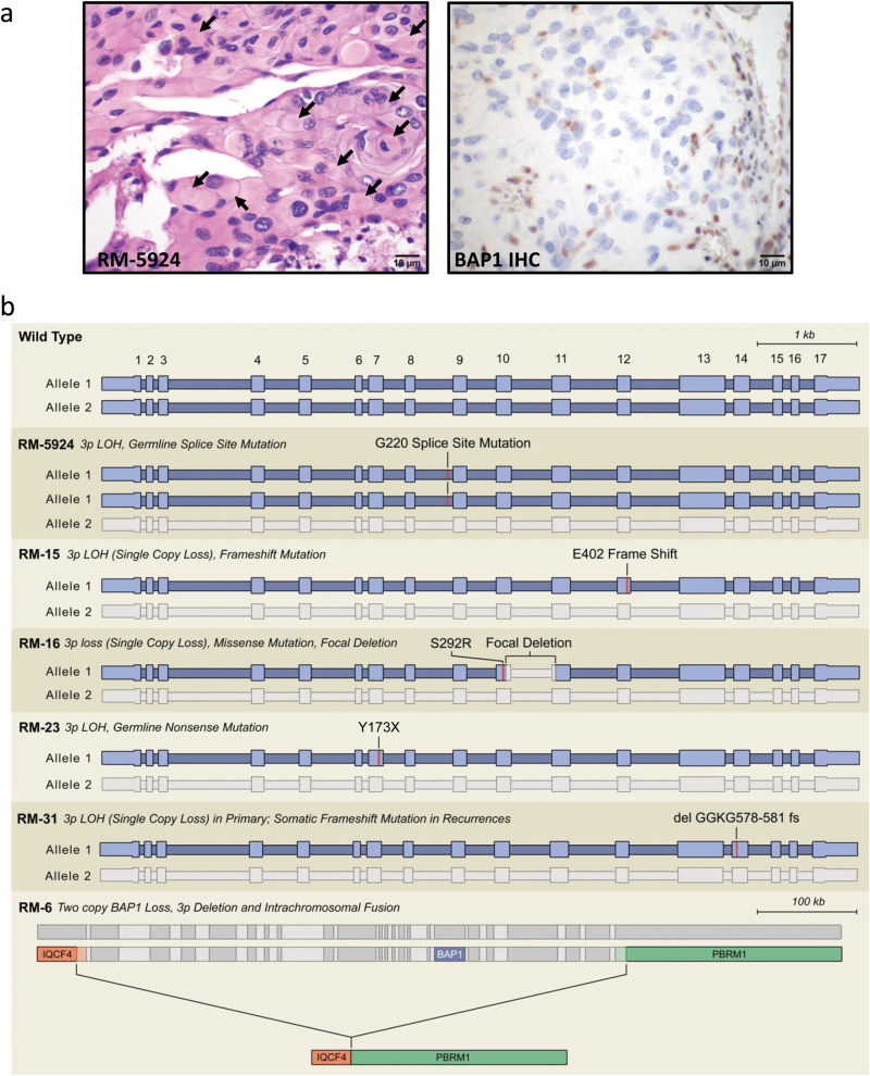
Methods: To define genomic aberrations of rhabdoid meningiomas, we performed sequencing of cancer-related genes in 27 meningiomas from 18 patients with rhabdoid features and evaluated breast cancer [BRCA]1-associated protein 1 (BAP1) expression by immunohistochemistry in 336 meningiomas. We assessed outcomes, germline status, and family history in patients with BAP1-negative rhabdoid meningiomas.

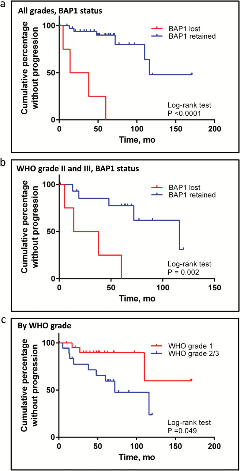
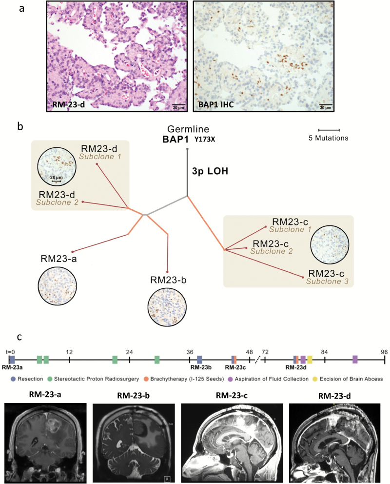
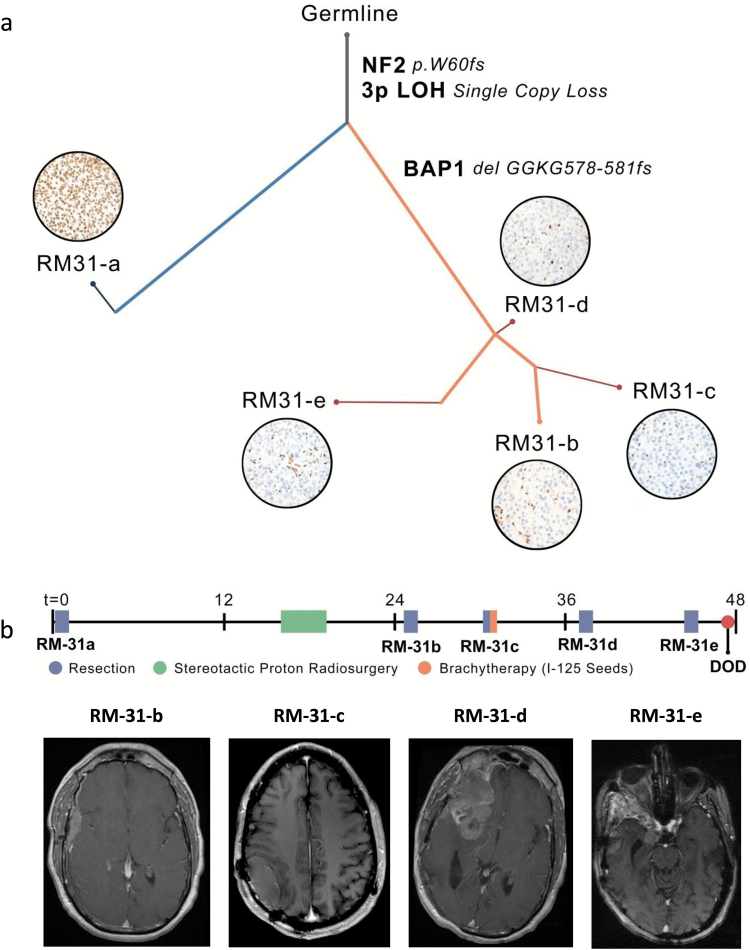
Results: The tumor suppressor gene BAP1, a ubiquitin carboxy-terminal hydrolase, is inactivated in a subset of high-grade rhabdoid meningiomas. Patients with BAP1-negative rhabdoid meningiomas had reduced time to recurrence compared with patients with BAP1-retained rhabdoid meningiomas (Kaplan-Meier analysis, 26 mo vs 116 mo, P < .001; hazard ratio 12.89). A subset of patients with BAP1-deficient rhabdoid meningiomas harbored germline BAP1 mutations, indicating that rhabdoid meningiomas can be a harbinger of the BAP1 cancer predisposition syndrome.

Conclusion: We define a subset of aggressive rhabdoid meningiomas that can be recognized using routine laboratory tests. We implicate ubiquitin deregulation in the pathogenesis of these high-grade malignancies. In addition, we show that familial and sporadic BAP1-mutated rhabdoid meningiomas are clinically aggressive, requiring intensive clinical management.

Keywords: BAP1; exome sequencing; rhabdoid meningiomas.


Figures

Fig. 1
Fig. 1
BAP1 loss in a high-grade rhabdoid meningioma. (A) Hematoxylin and eosin staining and BAP1 immunohistochemistry on sample RM-5924. Arrows highlight several of the globular paranuclear inclusions. (B) Schematic of BAP1 genetic aberrations resulting in BAP1 inactivation in syndromic and sporadic high-grade rhabdoid meningiomas. A list of the mutation calls made for these tumors is presented in Supplementary Tables 2, 6–8).
Fig. 2
Fig. 2
Kaplan–Meier survival analysis. Plot of the cumulative percentage without recurrence among the validation cohort of “meningioma with rhabdoid features” (BAP1 lost vs BAP1 retained) for patients with clinical follow-up harboring meningiomas of all grades (A) and only those with higher-grade (WHO II and III) meningiomas (B). Time to first progression used in analyses except if primary had documented intact BAP1 (eg, RM-31-a) in which time to progression after BAP1 loss is used (eg, time to RM-31-c from RM-31-b). Curves were compared using the log-rank (Mantel–Cox) test. P-value is displayed. Hazard ratio (HR) for BAP1-deficient tumors is listed in the text (log-rank): 12.9 when assessing all grades and 6.0 when assessing only higher grades (WHO grade II and III). (C) Plot of the cumulative percentage without recurrence among the validation cohort of WHO grade I meningioma and WHO grades II and grade III meningioma with rhabdoid features.
Fig. 3
Fig. 3
Characterization of genomic evolution of rhabdoid meningioma RM-23. (A) Co-occurrence of rhabdoid cytomorphology and papillary architecture in RM-23. (B) Phylogenetic tree inferred for familial rhabdoid meningioma RM-23 primary and recurrences. Branch thickness is proportional to the cancer-cell fraction (CCF) of mutations on that branch. Branch colors indicate types of tissue samples descended from each branch (gray, shared by all samples; blue, unique to primary sample; orange, present in recurrences; red, present in subclones of the recurrent tumor—eg, RM-23-c subclones 1, 2, and 3). Photomicrographs in circles show BAP1 immunohistochemistry for indicated tumors. Scale bar, 20µm. (C) Clinical information for patient RM-23. Timeline indicating relative sequence of major clinical events, including each surgery and other interventions. Representative images of preoperative MRI before each of the 4 surgical resections. While RM-23-a and RM-23-b were solitary masses, recurrences RM-23-c and RM-23-d comprised multiple spatially distinct masses along the falx. This pattern is consistent with the results from phylogenetic analysis showing that samples RM-23-c and RM-23-d comprised related but genetically distinct subclones. A list of mutation calls made for each of these 4 samples (RM-23-a to RM-23-d; noted as allelic fractions) is provided in Supplementary Table 7 (gene names and allelic fractions are in columns a–e).
Fig. 4
Fig. 4
Characterization of genomic evolution of rhabdoid meningioma RM-31. (A) Phylogenetic tree inferred for sporadic rhabdoid meningioma RM-31 primary and recurrences. Branch thickness is proportional to the cancer-cell fraction (CCF) of mutations on that branch. Branch colors indicate types of tissue samples descended from each branch (gray, shared by all samples; blue, unique to primary sample; orange, present in recurrences; red, present in subclones of the recurrent tumor—eg, RM-23-c subclones 1, 2, and 3). Photomicrographs in circles show BAP1 immunohistochemistry for the indicated tumor. Scale bar, 20µm. (B) Clinical information for patient RM-31. Timeline of treatment indicating the relative sequence of major clinical events, including each surgery and other treatment interventions. Representative images of MRI before the indicated surgical resections. MRI from the patient prior to resection of primary tumor (RM-31-a) was performed at an outside facility and images were unavailable. A list of mutation calls made for each of these 5 samples (RM-31-a to RM-31-e; noted as allelic fractions) is provided in Supplementary Table 8 (gene names and allelic fractions are in columns a–f).

References

    1. Louis DN, Ohgaki H, Wiestler OD, et al. WHO Classification of Tumours of the Central Nervous System. Revised 4th ed. Lyon, France: International Agency for Research on Cancer; 2016:232–257.
    1. Abedalthagafi MS, Merrill PH, Bi WL, et al. Angiomatous meningiomas have a distinct genetic profile with multiple chromosomal polysomies including polysomy of chromosome 5. Oncotarget. 2014;5(21):10596–10606. - PMC - PubMed
    1. Brastianos PK, Horowitz PM, Santagata S, et al. Genomic sequencing of meningiomas identifies oncogenic SMO and AKT1 mutations. Nat Genet. 2013;45(3):285–289. - PMC - PubMed
    1. Clark VE, Erson-Omay EZ, Serin A, et al. Genomic analysis of non-NF2 meningiomas reveals mutations in TRAF7, KLF4, AKT1, and SMO. Science. 2013;339(6123):1077–1080. - PMC - PubMed
    1. Reuss DE, Piro RM, Jones DT, et al. Secretory meningiomas are defined by combined KLF4 K409Q and TRAF7 mutations. Acta Neuropathol. 2013;125(3):351–358. - PubMed