. 2017 Jan;6(1):249-260.
doi: 10.5966/sctm.2016-0149. Epub 2016 Aug 29.

Genetic Modification of Human Peripheral Blood Aspirates Using Recombinant Adeno-Associated Viral Vectors for Articular Cartilage Repair with a Focus on Chondrogenic Transforming Growth Factor-β Gene Delivery

Affiliations

Genetic Modification of Human Peripheral Blood Aspirates Using Recombinant Adeno-Associated Viral Vectors for Articular Cartilage Repair with a Focus on Chondrogenic Transforming Growth Factor-β Gene Delivery

Janina Frisch et al. Stem Cells Transl Med. 2017 Jan.

Abstract

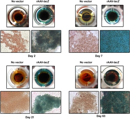
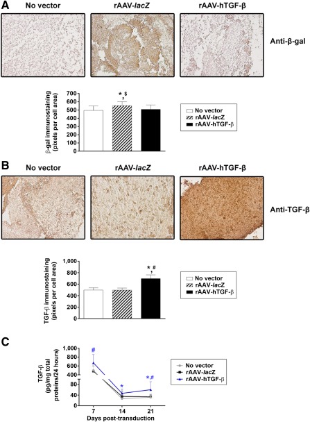
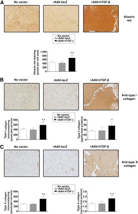
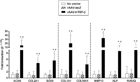
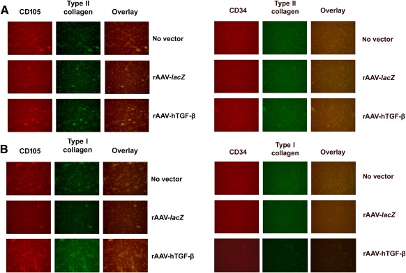
Transplantation of genetically modified peripheral blood aspirates that carry chondrogenically competent progenitor cells may offer new, convenient tools to treat articular cartilage lesions compared with the more complex and invasive application of bone marrow concentrates or of bone marrow-derived mesenchymal stem cells. Here, we show that recombinant adeno-associated viral (rAAV) vectors are powerful gene vehicles capable of successfully targeting primary human peripheral blood aspirates in a stable and safe manner, allowing for an efficient and long-term transgene expression in such samples (up to 63 days with use of a lacZ reporter gene and for at least 21 days with application of the pleiotropic, chondrogenic factor transforming growth factor-β [TGF-β]). rAAV-mediated overexpression of TGF-β enhanced both the proliferative and metabolic properties of the peripheral blood aspirates, also increasing the chondrogenic differentiation processes in these samples. Hypertrophy and osteogenic differentiation events were also activated by production of TGF-β via rAAV, suggesting that translation of the current approach in vivo will probably require close regulation of expression of this candidate gene. However, these results support the concept of directly modifying peripheral blood as a novel approach to conveniently treat articular cartilage lesions in patients. Stem Cells Translational Medicine 2017;6:249-260.

Keywords: Cartilage repair; Gene therapy; Peripheral blood aspirates; Transforming growth factor-β; rAAV vectors.


Figures

Figure 1
Figure 1
Detection of transgene expression in human peripheral blood aspirates after rAAV‐mediated gene transfer. Aspirates were transduced with rAAV‐lacZ or rAAV‐human transforming growth factor‐β (40 μl each vector) or left untreated, kept in regular growth medium, and processed to monitor transgene (lacZ) expression by X‐Gal staining at the denoted time points (original magnification, ×40; all representative data) as described in Materials and Methods. Abbreviation: rAAV, recombinant adeno‐associated viral.
Figure 2
Figure 2
Detection of transgene expression in chondrogenically induced human peripheral blood aspirates after rAAV‐mediated gene transfer. Aspirates were transduced with rAAV‐lacZ or rAAV‐hTGF‐β or left untreated (as described in Fig. 1), kept in chondrogenic medium for 21 days, and processed to monitor (A) lacZ expression by immunohistochemistry with corresponding histomorphometric analyses and (B, C) transforming growth factor‐β expression by immunohistochemistry with corresponding histomorphometric analyses (B) and enzyme‐linked immunosorbent assay (C) as described in Materials and Methods (original magnification, ×20; all representative data). ∗, statistically significant compared with no vector treatment; #, significant compared with rAAV‐lacZ; $, significant compared with rAAV‐hTGF‐β. Abbreviations: hTGF‐β, human transforming growth factor‐β; rAAV, recombinant adeno‐associated viral; TGF‐β, transforming growth factor‐β.
Figure 3
Figure 3
Evaluation of the proliferative activities in chondrogenically induced human peripheral blood aspirates upon rAAV‐mediated gene transfer. Aspirates were transduced with rAAV‐lacZ or rAAV‐hTGF‐β or left untreated (as described in Figs. 1 and 2), kept in chondrogenic medium for 21 days, and processed to evaluate (A) the cell densities on H&E‐stained histological sections with corresponding histomorphometric analyses (original magnification, ×20; representative data) and (B) the DNA contents as described in Materials and Methods. ∗, statistically significant compared with no vector treatment; #, significant compared with rAAV‐lacZ. Abbreviations: H&E, hematoxylin and eosin; hTGF‐β, human transforming growth factor‐β; rAAV, recombinant adeno‐associated viral; TGF‐β, transforming growth factor‐β.
Figure 4
Figure 4
Determination of the chondrogenic processes in chondrogenically induced human peripheral blood aspirates upon rAAV‐mediated gene transfer. Aspirates were transduced with rAAV‐lacZ or rAAV‐hTGF‐β or left untreated (as described in Figs. 1–3), kept in chondrogenic medium for 21 days, and processed for (A) toluidine blue staining with corresponding histomorphometric analyses and measurement of the proteoglycan contents, (B) type II collagen immunostaining with corresponding histomorphometric analyses and measurement of the type II collagen contents, and (C) SOX9 immunostaining with corresponding histomorphometric analyses, as described in Materials and Methods (original magnification, ×20; all representative data). ∗, Statistically significant compared with no vector treatment; #, significant compared with rAAV‐lacZ. Abbreviations: hTGF‐β, human transforming growth factor‐β; rAAV, recombinant adeno‐associated viral.
Figure 5
Figure 5
Evaluation of hypertrophic and terminal differentiation events in chondrogenically induced human peripheral blood aspirates upon rAAV‐mediated gene transfer. Aspirates were transduced with rAAV‐lacZ or rAAV‐hTGF‐β or left untreated (as described in Figs. 1–4), kept in chondrogenic medium for 21 days, and processed for (A) alizarin red staining with corresponding histomorphometric analyses, (B) type I collagen immunostaining with corresponding histomorphometric analyses and measurement of the type I collagen contents, and (C) type X collagen immunostaining with corresponding histomorphometric analyses and measurement of the type X collagen contents, as described in Materials and Methods (original magnification, ×20; all representative data). ∗, Statistically significant compared with no vector treatment; #, significant compared with rAAV‐lacZ. Abbreviations: hTGF‐β, human transforming growth factor‐β; rAAV, recombinant adeno‐associated viral.
Figure 6
Figure 6
Real‐time reverse transcriptase polymerase chain reaction analyses in chondrogenically induced human peripheral blood aspirates upon rAAV‐mediated gene transfer. Aspirates were transduced with rAAV‐lacZ or rAAV‐hTGF‐β or left untreated (as described in Figs. 1–5), kept in chondrogenic medium for 21 days, and analyzed for marker gene expression of ACAN, COL2A1, the transcription factor SOX9, COL1A1, COL10A1, MMP13, ALP, and the transcription factor RUNX2, with glyceraldehyde‐3‐phosphate dehydrogenase serving as a housekeeping gene and internal control for normalization. Ct values were generated for each target gene, and fold inductions (relative to untreated aspirates) were measured by using the 2−ΔΔCt method, as described in Materials and Methods. ∗, Statistically significant compared with no vector treatment; #, significant compared with rAAV‐lacZ. Abbreviations: ACAN, aggrecan; ALP, alkaline phosphatase; COL1A1, type I collagen; COL2A1, type II collagen; hTGF‐β, human transforming growth factor‐β; MMP13, matrix metalloproteinase 13; rAAV, recombinant adeno‐associated viral; RUNX2, runt‐related transcription factor 2.
Figure 7
Figure 7
Immunophenotypic analyses in chondrogenically induced human peripheral blood aspirates upon rAAV‐mediated gene transfer. Aspirates were transduced with rAAV‐lacZ or rAAV‐hTGF‐β or left untreated (as described in Figs. 1–6), kept in chondrogenic medium for 21 days, and analyzed for the coexpression of (A) CD105/type II collagen versus CD34/type II collagen and (B) CD105/type I collagen versus CD34/type I collagen by specific coimmunostaining, as described in Materials and Methods (original magnification, ×20; all representative data). Abbreviations: hTGF‐β, human transforming growth factor‐β; rAAV, recombinant adeno‐associated viral.

References

    1. Sophia Fox AJ, Bedi A, Rodeo SA The basic science of articular cartilage: Structure, composition, and function. Sports Health. 2009;1:461–468 - PMC - PubMed
    1. Buckwalter JA Articular cartilage: Injuries and potential for healing. J Orthop Sports Phys Ther. 1998;28:192–202 - PubMed
    1. Madry H, Grün UW, Knutsen G Cartilage repair and joint preservation: Medical and surgical treatment options. Dtsch Arztebl Int. 2011;108:669–677 - PMC - PubMed
    1. Zvaifler NJ, Marinova‐Mutafchieva L, Adams G et al. Mesenchymal precursor cells in the blood of normal individuals. Arthritis Res. 2000;2:477–488 - PMC - PubMed
    1. Chong PP, Selvaratnam L, Abbas AA et al. Human peripheral blood derived mesenchymal stem cells demonstrate similar characteristics and chondrogenic differentiation potential to bone marrow derived mesenchymal stem cells. J Orthop Res. 2012;30:634–642 - PubMed

Publication types

Substances

LinkOut - more resources